

A Unique Acylated Flavonol Glycoside from Prunus persica (L.) var. Florida Prince: A New Solid Lipid Nanoparticle Cosmeceutical Formulation for Skincare
- PMID: 33809166
- PMCID: PMC7998748
- DOI: 10.3390/antiox10030436
A Unique Acylated Flavonol Glycoside from Prunus persica (L.) var. Florida Prince: A New Solid Lipid Nanoparticle Cosmeceutical Formulation for Skincare
Abstract
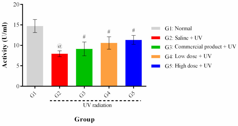
Polyphenols are known dietary antioxidants. They have recently attracted considerable interest in uses to prevent skin aging and hyperpigmentation resulting from solar UV-irradiation. Prunus persica (L.) leaves are considered by-products and were reported to have a remarkable antioxidant activity due to their high content of polyphenols. This study aimed at the development of a cosmeceutical anti-aging and skin whitening cream preparation using ethanol leaves extract of Prunus persica (L.) (PPEE) loaded in solid lipid nanoparticles (SLNs) to enhance the skin delivery. Chemical investigation of PPEE showed significantly high total phenolic and flavonoids content with notable antioxidant activities (DPPH, ABTS, and β-carotene assays). A unique acylated kaempferol glycoside with a rare structure, kaempferol 3-O-β-4C1-(6″-O-3,4-dihydroxyphenylacetyl glucopyranoside) (KDPAG) was isolated for the first time and its structure fully elucidated. It represents the first example of acylation with 3,4-dihydroxyphenyl acetic acid in flavonoid chemistry. The in-vitro cytotoxicity studies against a human keratinocytes cell line revealed the non-toxicity of PPEE and PPEE-SLNs. Moreover, PPEE, PPEE-SLNs, and KDPAG showed good anti-elastase activity, comparable to that of N-(Methoxysuccinyl)-Ala-Ala-Pro-Val-chloromethyl ketone. Besides, PPEE-SLNs and KDPAG showed significantly (p < 0.001) higher anti-collagenase and anti-tyrosinase activities in comparison to EDTA and kojic acid, respectively. Different PPEE-SLNs cream formulae (2% and 5%) were evaluated for possible anti-wrinkle activity against UV-induced photoaging in a mouse model using a wrinkle scoring method and were shown to offer a highly significant protective effect against UV, as evidenced by tissue biomarkers (SOD) and histopathological studies. Thus, the current study demonstrates that Prunus persica leaf by-products provide an interesting, valuable resource for natural cosmetic ingredients. This provides related data for further studying the potential safe use of PPEE-SLNs in topical anti-aging cosmetic formulations with enhanced skin permeation properties.
Keywords: antioxidant; in-vitro skin related enzymes; in-vivo anti-wrinkle; kaempferol 3-O-β-4C1-(6″-O-3,4-dihydroxyphenylacetyl glucopyranoside) by-products; solid lipid nanoparticles.
Conflict of interest statement
The authors declare no conflict of interest.
Figures


References
-
- Jiratchayamaethasakul C., Ding Y., Hwang O., Im S.-T., Jang Y., Myung S.-W., Lee J.M., Kim H.-S., Ko S.-C., Lee S.-H. In vitro screening of elastase, collagenase, hyaluronidase, and tyrosinase inhibitory and antioxidant activities of 22 halophyte plant extracts for novel cosmeceuticals. Fish. Aquat. Sci. 2020;23:1–9. doi: 10.1186/s41240-020-00149-8. - DOI
-
- Hwang I.S., Kim J.E., Choi S.I., Lee H.R., Lee Y.J., Jang M.J., Son H.J., Lee H.S., Oh C.H., Kim B.H. UV radiation-induced skin aging in hairless mice is effectively prevented by oral intake of sea buckthorn (Hippophae rhamnoides L.) fruit blend for 6 weeks through MMP suppression and increase of SOD activity. Int. J. Mol. Med. 2012;30:392–400. doi: 10.3892/ijmm.2012.1011. - DOI - PubMed
-
- Garg C. Molecular mechanisms of skin photoaging and plant inhibitors. Int. J. Green Pharm. 2017;11:3268.
-
- Kang M., Park S.-H., Oh S.W., Lee S.E., Yoo J.A., Nho Y.H., Lee S., Han B.S., Cho J.Y., Lee J. Anti-melanogenic effects of resorcinol are mediated by suppression of cAMP signaling and activation of p38 MAPK signaling. Biosci. Biotechnol. Biochem. 2018;82:1188–1196. doi: 10.1080/09168451.2018.1459176. - DOI - PubMed
LinkOut - more resources
Full Text Sources
Other Literature Sources
