. 2021 Mar 12;21(6):2020.
doi: 10.3390/s21062020.

Effect of a Brain-Computer Interface Based on Pedaling Motor Imagery on Cortical Excitability and Connectivity

Affiliations

Effect of a Brain-Computer Interface Based on Pedaling Motor Imagery on Cortical Excitability and Connectivity

Vivianne Flávia Cardoso et al. Sensors (Basel). .

Abstract

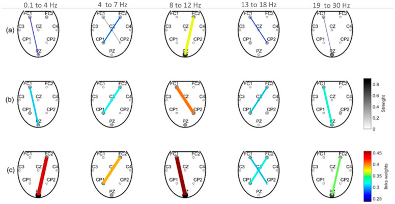
Recently, studies on cycling-based brain-computer interfaces (BCIs) have been standing out due to their potential for lower-limb recovery. In this scenario, the behaviors of the sensory motor rhythms and the brain connectivity present themselves as sources of information that can contribute to interpreting the cortical effect of these technologies. This study aims to analyze how sensory motor rhythms and cortical connectivity behave when volunteers command reactive motor imagery (MI) BCI that provides passive pedaling feedback. We studied 8 healthy subjects who performed pedaling MI to command an electroencephalography (EEG)-based BCI with a motorized pedal to receive passive movements as feedback. The EEG data were analyzed under the following four conditions: resting, MI calibration, MI online, and receiving passive pedaling (on-line phase). Most subjects produced, over the foot area, significant event-related desynchronization (ERD) patterns around Cz when performing MI and receiving passive pedaling. The sharpest decrease was found for the low beta band. The connectivity results revealed an exchange of information between the supplementary motor area (SMA) and parietal regions during MI and passive pedaling. Our findings point to the primary motor cortex activation for most participants and the connectivity between SMA and parietal regions during pedaling MI and passive pedaling.

Keywords: brain connectivity; brain–computer interface; lower limb rehabilitation; motor sensory rhythms; pedaling.


Conflict of interest statement

On behalf of all authors, the corresponding author states that there is no conflict of interest.

Figures

Figure 1
Figure 1
Experimental setup using our brain–computer interface scheme.
Figure 2
Figure 2
Sequence followed during the experimental protocol.
Figure 3
Figure 3
The experimental paradigm: (a) calibration phase, and (b) online phase.
Figure 4
Figure 4
(a) Average relative power computed on 8 healthy subjects for three states, where pc and p-values compare the motor imagery (MI) calibration phase, online phase, and passive pedaling conditions in the online phase; (b) comparison of relative power over the Cz location analyzing the contribution of each frequency band when participants executed four tasks: rest state, MI during the calibration phase, instantly triggering the brain–computer interface (BCI) by MI, and receiving passive pedaling.
Figure 5
Figure 5
Significant event-related desynchronization (ERD) analysis. (a) Distribution of significant ERD changes in the time–frequency representation over Cz, and topographic maps of mean ERD power for the mu band, and the low and high beta bands, obtained when participants executed MI in the calibration phase and online phases. (b) Significant ERD power average for subject-specific bands and latency of ERD peaks during MI and passive pedaling in closed-loop.
Figure 6
Figure 6
(a) Representation of significant ERD patterns using the time–frequency representation, where the intervals from −1.0 to 0 s and from 0 to 4.5 s are respectively related to MI before triggering the BCI and passive pedaling. (b) Average of ERD peaks for subject-specific frequency bands during MI and passive pedaling in closed-loop.
Figure 7
Figure 7
Connectivity between surface cortical areas considering flow and force between the electroencephalography (EEG) channels for the delta, theta, mu band, and beta bands, during three conditions: (a) MI calibration, (b) MI online, and (c) passive movements.

References

    1. Bastos-Filho T.F. Introduction to Non-Invasive EEG-Based Brain-Computer Interfaces for Assistive Technologies. CRC Press; Boca Raton, FL, USA: 2020.
    1. Takeuchi N., Izumi S.-I. Rehabilitation with Poststroke Motor Recovery: A Review with a Focus on Neural Plasticity. Stroke Res. Treat. 2013;2013:1–13. doi: 10.1155/2013/128641. - DOI - PMC - PubMed
    1. Li M., Xu G., Xie J., Chen C. A review: Motor rehabilitation after stroke with control based on human intent. Proc. Inst. Mech. Eng. Part H: J. Eng. Med. 2018;232:344–360. doi: 10.1177/0954411918755828. - DOI - PubMed
    1. Liu D., Chen W., Lee K., Pei Z., Millan J.D.R. An EEG-based brain-computer interface for gait training; Proceedings of the 2017 29th Chinese Control and Decision Conference (CCDC); Chongqing, China. 28–30 May 2017; pp. 6755–6760.
    1. Hashimoto Y., Ushiba J. EEG-based classification of imaginary left and right foot movements using beta rebound. Clin. Neurophysiol. 2013;124:2153–2160. doi: 10.1016/j.clinph.2013.05.006. - DOI - PubMed

LinkOut - more resources