

The Discovery of Endoplasmic Reticulum Storage Disease. The Connection between an H&E Slide and the Brain
- PMID: 33809321
- PMCID: PMC8001541
- DOI: 10.3390/ijms22062899
The Discovery of Endoplasmic Reticulum Storage Disease. The Connection between an H&E Slide and the Brain
Abstract
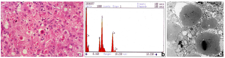
The revolutionary evolution in science and technology over the last few decades has made it possible to face more adequately three main challenges of modern medicine: changes in old diseases, the appearance of new diseases, and diseases that are unknown (mostly genetic), despite research efforts. In this paper we review the road travelled by pathologists in search of a method based upon the use of routine instruments and techniques which once were available for research only. The application to tissue studies of techniques from immunology, molecular biology, and genetics has allowed dynamic interpretations of biological phenomena with special regard to gene regulation and expression. That implies stepwise investigations, including light microscopy, immunohistochemistry, in situ hybridization, electron microscopy, molecular histopathology, protein crystallography, and gene sequencing, in order to progress from suggestive features detectable in routinely stained preparations to more characteristic, specific, and finally, pathognomonic features. Hematoxylin and Eosin (H&E)-stained preparations and appropriate immunohistochemical stains have enabled the recognition of phenotypic changes which may reflect genotypic alterations. That has been the case with hepatocytic inclusions detected in H&E-stained preparations, which appeared to correspond to secretory proteins that, due to genetic mutations, were retained within the rough endoplasmic reticulum (RER) and were deficient in plasma. The identification of this phenomenon affecting the molecules alpha-1-antitrypsin and fibrinogen has led to the discovery of a new field of cell organelle pathology, endoplasmic reticulum storage disease(s) (ERSD). Over fifty years, pathologists have wandered through a dark forest of complicated molecules with strange conformations, and by detailed observations in simple histopathological sections, accompanied by a growing background of molecular techniques and revelations, have been able to recognize and identify arrays of grotesque polypeptide arrangements.
Keywords: alpha-1-antitrypsin deficiency; endoplasmic reticulum storage disease; hereditary hypofibrinogenemia hepatic storage.
Conflict of interest statement
The authors declare no conflict of interest.
Figures


Similar articles
-
Hepatic and Extrahepatic Sources and Manifestations in Endoplasmic Reticulum Storage Diseases.Int J Mol Sci. 2021 May 28;22(11):5778. doi: 10.3390/ijms22115778. Int J Mol Sci. 2021. PMID: 34071368 Free PMC article. Review.
-
The Recruitment-Secretory Block ("R-SB") Phenomenon and Endoplasmic Reticulum Storage Diseases.Int J Mol Sci. 2021 Jun 24;22(13):6807. doi: 10.3390/ijms22136807. Int J Mol Sci. 2021. PMID: 34202771 Free PMC article. Review.
-
From immunohistochemistry to in situ hybridization.Liver. 1992 Aug;12(4 Pt 2):290-5. doi: 10.1111/j.1600-0676.1992.tb01063.x. Liver. 1992. PMID: 1447961 Review.
-
Hepatic endoplasmic reticulum storage diseases.Liver. 1992 Dec;12(6):357-62. doi: 10.1111/j.1600-0676.1992.tb00589.x. Liver. 1992. PMID: 1470006 Review.
-
Mineralization of alpha-1-antitrypsin inclusion bodies in Mmalton alpha-1-antitrypsin deficiency.Orphanet J Rare Dis. 2018 May 16;13(1):79. doi: 10.1186/s13023-018-0821-7. Orphanet J Rare Dis. 2018. PMID: 29769092 Free PMC article.
Cited by
-
Hepatic and Extrahepatic Sources and Manifestations in Endoplasmic Reticulum Storage Diseases.Int J Mol Sci. 2021 May 28;22(11):5778. doi: 10.3390/ijms22115778. Int J Mol Sci. 2021. PMID: 34071368 Free PMC article. Review.
-
Protein Aggregation in the ER: Calm behind the Storm.Cells. 2021 Nov 28;10(12):3337. doi: 10.3390/cells10123337. Cells. 2021. PMID: 34943844 Free PMC article. Review.
-
The Recruitment-Secretory Block ("R-SB") Phenomenon and Endoplasmic Reticulum Storage Diseases.Int J Mol Sci. 2021 Jun 24;22(13):6807. doi: 10.3390/ijms22136807. Int J Mol Sci. 2021. PMID: 34202771 Free PMC article. Review.
-
A missense variant in DGKG as a recessive functional variant for hepatic fibrinogen storage disease in Wagyu cattle.J Vet Intern Med. 2023 Nov-Dec;37(6):2631-2637. doi: 10.1111/jvim.16865. Epub 2023 Sep 8. J Vet Intern Med. 2023. PMID: 37681469 Free PMC article.
-
Protein quality control and aggregation in the endoplasmic reticulum: From basic to bedside.Front Cell Dev Biol. 2023 Apr 19;11:1156152. doi: 10.3389/fcell.2023.1156152. eCollection 2023. Front Cell Dev Biol. 2023. PMID: 37152279 Free PMC article. Review.
References
-
- Sharp H.L., Bridges B.A., Krivit W., Freier E.F. Cirrhosis associated with alpha-1-antitrypsin deficiency—A previous unrecognized inheritd disorder. J. Lab. Clin. Med. 1969;73:934–939. - PubMed
-
- Callea F., De Vos R., Pinackat J., Favret M., Facchetti F., Fiaccavento S., Ascari E., Tortora O., Albertini A., Henschen A., et al. Hereditary hypofinogenemia with hepatic storage of fibrinogen: A new endoplasmic reticulum storage diseases. In: Lowe G.D.O., editor. Fibrinogen 2, Biochemistry, Physiology and Clinical Relevance. Elsevier; Amsterdam, The Netherlands: 1987. pp. 75–78.
-
- Majno G., Joris I. Cells Tissues and Disease. Oxford University Press; Oxford, UK: 2004.
-
- Ishak K.G., Sharp H.L., Schwarzenberg S.J. Metabolic Errors and Liver Disease. In: RNM, editor. Pathology of the Liver. Churchill Livingstone; London, UK: 2003.
Publication types
MeSH terms
Substances
LinkOut - more resources
Full Text Sources
Other Literature Sources
Medical
