. 2021 Mar 16;10(3):460.
doi: 10.3390/antiox10030460.

Rolipram Prevents the Formation of Abdominal Aortic Aneurysm (AAA) in Mice: PDE4B as a Target in AAA

Affiliations

Rolipram Prevents the Formation of Abdominal Aortic Aneurysm (AAA) in Mice: PDE4B as a Target in AAA

Saray Varona et al. Antioxidants (Basel). .

Abstract

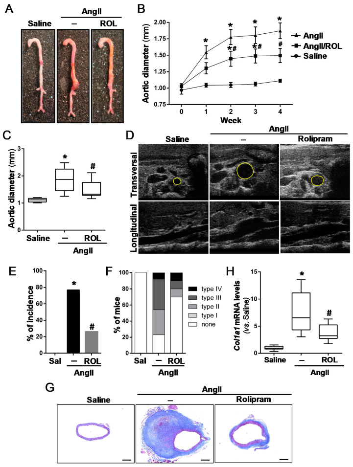
Abdominal aortic aneurysm (AAA) is a common life-threatening condition characterized by exacerbated inflammation and the generation of reactive oxygen species. Pharmacological treatments to slow AAA progression or to prevent its rupture remain a challenge. Targeting phosphodiesterase 4 (PDE4) has been verified as an effective therapeutic strategy for an array of inflammatory conditions; however, no studies have assessed yet PDE4 in AAA. Here, we used angiotensin II (AngII)-infused apolipoprotein E deficient mice to study the involvement of the PDE4 subfamily in aneurysmal disease. PDE4B but not PDE4D was upregulated in inflammatory cells from both experimental and human AAA. The administration of the PDE4 selective inhibitor rolipram (3 mg/kg/day) to AngII-challenged mice (1000 ng/kg bodyweight/min) protected against AAA formation, limiting the progressive increase in the aortic diameter without affecting the blood pressure. The drug strongly attenuated the rise in vascular oxidative stress (superoxide anion) induced by AngII, and decreased the expression of inflammatory markers, as well as the recruitment of macrophages (MAC3+), lymphocytes (CD3+), and neutrophils (ELANE+) into the vessel wall. Rolipram also normalized the vascular MMP2 expression and MMP activity, preserving the elastin integrity and improving the vascular remodelling. These results point to PDE4B as a new therapeutic target for AAA.

Keywords: PDE4B; abdominal aortic aneurysm; reactive oxygen species; rolipram.


Conflict of interest statement

The authors declare no conflict of interest.

Figures

Figure 1
Figure 1
PDE4B expression is up-regulated in the abdominal aortas from AngII-infused ApoE−/− mice. ApoE−/− mice were infused with AngII (1000 ng/kg/min) or saline solution for 28 days. (A) The PDE4B expression in abdominal aortas from these animals was assessed by Western blot. The levels of β-actin are shown as a loading control. The boxplot on the right shows the quantification of the PDE4B protein levels. The box extends from the 25th to the 75th percentile, and the median is indicated by a horizontal line. The whiskers represent the maximum and minimum values (Saline, n = 4; Ang II n = 6); * p < 0.01 vs. saline. (B) Representative PDE4B immunostaining in these samples. The arrowheads indicate the PD4B-positive cells in aneurysmal tissues. Bars: 20 µm.
Figure 2
Figure 2
PDE4B expression is enhanced in human AAA. (A) PDE4B expression in abdominal aorta from AAA patients (AAA) and healthy donors (Do), assessed by real-time PCR. The data are presented as boxplots. The box extends from the 25th to the 75th percentile, and the median is indicated by a horizontal line. The whiskers represent the maximum and minimum values (DO, n = 14; AAA n = 61); * p < 0.001 vs. Donors. (B) Representative PDE4B immunostaining in healthy aorta from donors and aneurysmal tissues from AAA patients (top panels; bar: 100 µm). The boxed areas are shown at a higher magnification below, and the arrowheads indicate PDE4B-positive cells (lower panels; bar: 50 µm). (C) PDE4D mRNA levels in these samples.
Figure 3
Figure 3
Rolipram ameliorates AAA development in AngII-infused ApoE−/− mice. ApoE−/− mice were infused with saline solution (Sal) or AngII (1000 ng/kg/min) for 28 days. The mice challenged with AngII were treated or not with rolipram (ROL, 3 mg/kg/day). (A) Representative macroscopic images of the aortas from each experimental group. (B) Time-course analysis of the abdominal aortic diameter by ecochardiography. The data are mean ± SEM (saline-infused mice, n = 9; AngII-infused animals, n = 13; AngII-infused mice treated with rolipram, n = 10). p < 0.05: * vs. saline-infused mice; # vs. AngII-challenged mice. (C) Quantitative analysis of the aortic diameter at the end of the experimental period (28 days). The results are expressed as boxplots. The box extends from the 25th to the 75th percentile, and the median is indicated by a horizontal line. The whiskers represent the maximum and minimum values. p < 0.05: * vs. saline-infused mice at the same time; # vs. AngII-challenged mice. (D) Representative ultrasonographic frames after 28 days of AngII infusion. Transverse and longitudinal images taken at the level of the suprarenal aorta are shown. The aortic diameter is indicated with a yellow line. (E) Incidence of AAA in each experimental group. p < 0.05: * vs. saline-infused mice; #vs. AngII-challenged mice. (F) The aneurysm severity based on the Manning scale. (G) Masson trichrome staining of the abdominal aortas from each group. Representative images are shown. Bars: 200 µm. (H) The Col1A1 mRNA levels assessed in these samples by real-time PCR. The data are presented as box and whisker plots. The box extends from the 25th to the 75th percentile, and the median is indicated by a horizontal line. The whiskers represent the maximum and minimum values (saline-infused mice n = 8; AngII-challenged mice, n = 12; AngII-challenged mice treated with rolipram, n = 8). p < 0.05; * vs. saline-infused mice; # vs. AngII-challenged mice.
Figure 4
Figure 4
Inflammatory cell infiltration in the abdominal aorta from AngII-infused ApoE−/− mice is attenuated by rolipram. ApoE−/− mice were infused with saline solution or AngII (1000 ng/kg/min) for 28 days. The mice challenged with AngII were treated or not with rolipram (3 mg/kg/day). (AC) Representative images corresponding to macrophage ((A) MAC3), lymphocyte ((B) CD3) and neutrophil ((C) neutrophil elastase, ELANE) infiltration, analysed by immunohistochemistry. The arrowheads indicate positive cells (Bars: 50 µm). The boxplots on the right show the quantification of the positive cells for Mac-3, CD3, and the neutrophil elastase per aortic section. The box extends from the 25th to the 75th percentile, and the median is indicated by a horizontal line. The whiskers represent the maximum and minimum values (saline-infused mice, n = 5; AngII-challenged mice, n = 8; AngII-challenged mice treated with rolipram, n = 6). In (AC), p < 0.05: * vs. saline-infused mice; # vs. AngII-challenged mice.
Figure 5
Figure 5
Rolipram limits the inflammatory response triggered by AngII in ApoE−/− mice. ApoE−/− mice were infused with saline solution or AngII (1000 ng/kg/min) for 28 days. Mice challenged with AngII were treated or not with rolipram (3 mg/kg/day). (A,B) The Emr1 (A) and Mcp1 (B) mRNA levels were analysed by real-time PCR (saline-infused mice n = 8; AngII-challenged mice, n = 12; AngII-challenged mice treated with rolipram, n = 9). (C) The immunohistochemical analysis of MCP1 expression in the abdominal aortas from each experimental group. Representative images are shown (Bar: 50 µm; saline-infused mice n = 5; AngII-challenged mice, n = 8; AngII-challenged mice treated with rolipram, n = 6). The data are presented as box and whisker plots. The box extends from the 25th to the 75th percentile, and the median is indicated by a horizontal line. The whiskers represent the maximum and minimum values. In (AC), p < 0.05: * vs. saline-infused mice; # vs. AngII-challenged mice.
Figure 6
Figure 6
Rolipram reduces the increased production of vascular superoxide anions induced by AngII in ApoE−/− mice. The ApoE−/− mice were infused with saline solution or AngII (1000 ng/kg/min) for 28 days. The mice challenged with AngII were treated or not with rolipram (3 mg/kg/day). Their vascular superoxide anion production was assessed by DHE staining in the abdominal aorta sections of each experimental group. Representative images of the DHE staining are shown (Bars: 50 µm). The data are presented as box and whisker plots. The box extends from the 25th to the 75th percentile, and median is indicated by a horizontal line. The whiskers represent the maximum and minimum values (saline-infused mice n = 6; AngII-challenged mice, n = 8; AngII-challenged mice treated with rolipram, n = 7). p < 0.01: * vs. saline-infused mice; # vs. AngII-challenged mice.
Figure 7
Figure 7
Rolipram attenuates the vascular remodelling induced by AngII in ApoE−/− mice. ApoE−/− mice were infused with saline solution or AngII (1000 ng/kg/min) for 28 days. The mice challenged with AngII were treated or not with rolipram (3 mg/kg/day). (A) Representative images showing the structure of the elastin fibres in the abdominal aortas after orcein staining. The arrowheads indicate elastin ruptures (Bars: 100 µm). (B) The boxplot shows the quantitative analysis of the number of ruptures in the elastin fibers per aortic section. The box extends from the 25th to the 75th percentile, and the median is indicated by a horizontal line. The whiskers represent the maximum and minimum values (saline-infused mice n = 6; AngII-challenged mice, n = 12; AngII-challenged mice treated with rolipram, n = 7). p < 0.05: * vs. saline-infused mice; # vs. AngII-challenged mice.
Figure 8
Figure 8
Rolipram attenuates the increase in MMP activity triggered by AngII in ApoE−/− mice. ApoE−/− mice were infused with saline solution or AngII (1000 ng/kg/min) for 28 days. The mice challenged with AngII were treated or not with rolipram (3 mg/kg/day). (A,B) The Mmp2 (A) and Mmp9 (B) mRNA levels were analysed by real-time PCR. The data are presented as box and whisker plots. The box extends from the 25th to the 75th percentile, and the median is indicated by a horizontal line. The whiskers represent the maximum and minimum values (saline-infused mice n = 8; AngII-challenged mice, n = 12; AngII-challenged mice treated with rolipram, n = 9). (C) The MMP activity analysed by in situ zymography in the abdominal aortas from each experimental group. Representive images are shown (Bars: 50 µm). The boxplot on the right shows the quantification of the MMP activity (saline-infused mice n = 5; AngII-challenged mice, n = 6; AngII-challenged mice treated with rolipram, n = 6). In A to C, p < 0.05: * vs. saline-infused mice; # vs. AngII-challenged mice.

Similar articles

Cited by

References

    1. Nordon I.M., Hinchliffe R.J., Loftus I.M., Thompson M.M. Pathophysiology and epidemiology of abdominal aortic aneurysms. Nat. Rev. Cardiol. 2011;8:92–102. doi: 10.1038/nrcardio.2010.180. - DOI - PubMed
    1. Kent K.C. Clinical practice. Abdominal aortic aneurysms. N. Engl. J. Med. 2014;371:2101–2108. doi: 10.1056/NEJMcp1401430. - DOI - PubMed
    1. GBD 2013 Mortality and Causes of Death Collaborators Global, regional, and national age-sex specific all-cause and cause-specific mortality for 240 causes of death, 1990–2013: A systematic analysis for the Global Burden of Disease Study 2013. Lancet. 2015;385:117–171. doi: 10.1016/S0140-6736(14)61682-2. - DOI - PMC - PubMed
    1. Golledge J. Abdominal aortic aneurysm: Update on pathogenesis and medical treatments. Nat. Rev. Cardiol. 2019;16:225–242. doi: 10.1038/s41569-018-0114-9. - DOI - PubMed
    1. Chaikof E.L., Dalman R.L., Eskandari M.K., Jackson B.M., Lee W.A., Mansour M.A., Mastracci T.M., Mell M., Murad M.H., Nguyen L.L., et al. The Society for Vascular Surgery practice guidelines on the care of patients with an abdominal aortic aneurysm. J. Vasc. Surg. 2018;67:2–77.e2. doi: 10.1016/j.jvs.2017.10.044. - DOI - PubMed