Review
. 2021 Mar 16;22(6):3013.
doi: 10.3390/ijms22063013.

Central Apparatus, the Molecular Kickstarter of Ciliary and Flagellar Nanomachines

Affiliations
Review

Central Apparatus, the Molecular Kickstarter of Ciliary and Flagellar Nanomachines

Zuzanna Samsel et al. Int J Mol Sci. .

Abstract

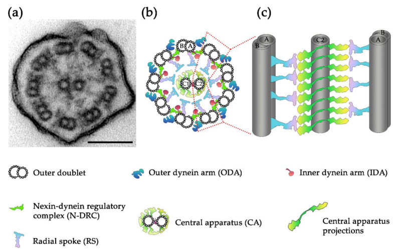
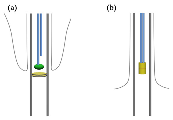
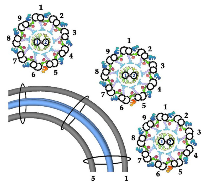
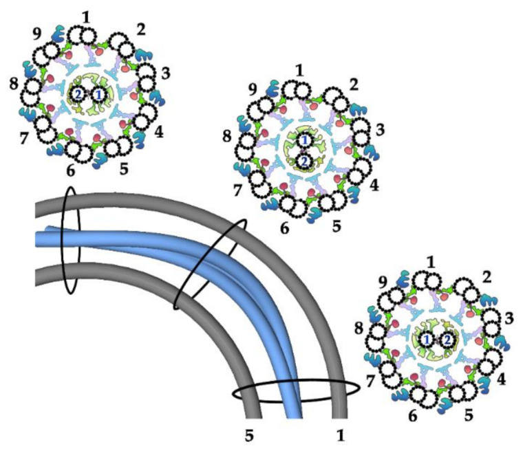
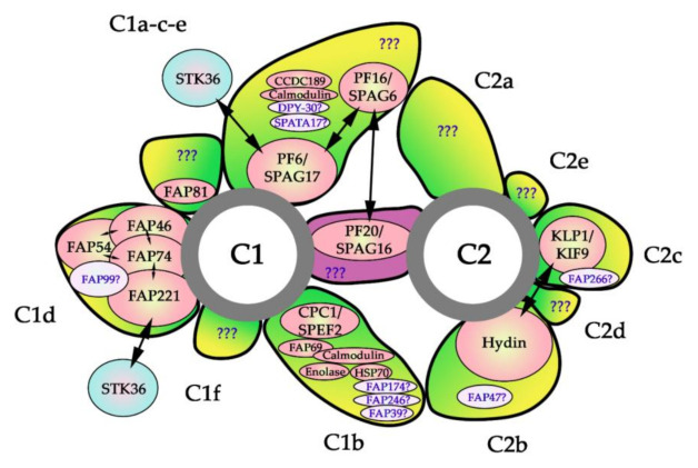
Motile cilia and homologous organelles, the flagella, are an early evolutionarily invention, enabling primitive eukaryotic cells to survive and reproduce. In animals, cilia have undergone functional and structural speciation giving raise to typical motile cilia, motile nodal cilia, and sensory immotile cilia. In contrast to other cilia types, typical motile cilia are able to beat in complex, two-phase movements. Moreover, they contain many additional structures, including central apparatus, composed of two single microtubules connected by a bridge-like structure and assembling numerous complexes called projections. A growing body of evidence supports the important role of the central apparatus in the generation and regulation of the motile cilia movement. Here we review data concerning the central apparatus structure, protein composition, and the significance of its components in ciliary beating regulation.

Keywords: Chlamydomonas; PCD; Trypanosoma; axoneme; central pair microtubules; male infertility.


Conflict of interest statement

The authors declare no conflict of interest.

Figures

Figure 1
Figure 1
Structure of motile cilia/flagella (CFs): (a) a transmission electron microscopy (TEM) cross-section of a Tetrahymena thermophila cilium, bar = 100 nm; (b) the organization of axonemal complexes across the CF circumference, C1 and C2—central microtubules 1 and 2, respectively, A and B—tubules A and B of the outer doublet; (c) the longitudinal organization of the outer doublet radial spokes (RS) and central apparatus (CA) projections showing possible interactions between these structures; for the simplicity only C2 microtubule is shown. Note that for clarity, other outer doublet complexes (ODA, IDA and N-DRC) are not shown.
Figure 2
Figure 2
Docking of the CA microtubule minus-end: (a,b) a schematic representation of the basal body and proximal part of the cilium in a ciliate and green alga (a) in ciliates only one of CA microtubules is attached to the axosome (green oval), while the other starts more distally, yellow discs represents plates of the transition zone [46]; (b) both central microtubules are attached in the tube/central cylinder (yellow tube) in Chlamydomonas [24].
Figure 3
Figure 3
Fixed position of CA microtubules in Strongylocentrotus CF: scheme showing bent CF in longitudinal view with outer doublets number 1 and 5 (gray) and CA microtubules (blue) and in three cross-sections: before, at and after the bend (as shown on the longitudinal view of the cilium). Note that CA microtubules run parallel to each other along the entire cilium length and that their position is constant in relation to the outer doublets (each projection interacts with RS of the same outer doublet at the whole CF length). The position of the outer doublets and CA with respect to the bend, according to [60].
Figure 4
Figure 4
Twisted position of CA microtubules in Chlamydomonas CF: scheme showing bent CF in longitudinal view with outer doublets number 1 and 5 (gray) and CA microtubules (blue) and in three cross-sections: before, at and after the bend (as shown on the longitudinal view of the CF). Note that position of the CA in relation to the outer doublets changes (each projection interacts with RS of different outer doublets, depending on the position in relation to the bend). The position of the outer doublets and CA with respect to the bend, according to [60].
Figure 5
Figure 5
Predicted localization of the evolutionarily conserved subunits of the CA. C1 and C2 —central microtubules 1 and 2, respectively. The experimentally confirmed CA components are represented by peach ovals, putative subunits are shown as light pink ovals. Blue ovals represent protein transiently or weakly associated with CA; two-way arrows mark experimentally confirmed interactions between CA subunits. The question marks in the bridge and projections indicate putative unidentified subunits. See the text and Table 1 for details.

Similar articles

Cited by

References

    1. Carvalho-Santos Z., Azimzadeh J., Pereira-Leal J.B., Bettencourt-Dias M. Tracing the Origins of Centrioles, Cilia, and Flagella. J. Cell Biol. 2011;194:165–175. doi: 10.1083/jcb.201011152. - DOI - PMC - PubMed
    1. Mitchell D.R. Evolution of Cilia. Cold Spring Harb. Perspect. Biol. 2017;9 doi: 10.1101/cshperspect.a028290. - DOI - PMC - PubMed
    1. Brightman M.W., Palay S.L. The fine structure of ependyma in the brain of the rat. J. Cell Biol. 1963;19:415–439. doi: 10.1083/jcb.19.2.415. - DOI - PMC - PubMed
    1. Breeze R.G., Wheeldon E.B. The Cells of the Pulmonary Airways. Am. Rev. Respir. Dis. 1977;116:705–777. doi: 10.1164/arrd.1977.116.4.705. - DOI - PubMed
    1. Legendre M., Zaragosi L.E., Mitchison H.M. Motile Cilia and Airway Disease. Semin. Cell Dev. Biol. 2020 doi: 10.1016/j.semcdb.2020.11.007. - DOI - PubMed

LinkOut - more resources