

Accuracy of Computer-Assisted Surgery in Maxillary Reconstruction: A Systematic Review
- PMID: 33809600
- PMCID: PMC8002284
- DOI: 10.3390/jcm10061226
Accuracy of Computer-Assisted Surgery in Maxillary Reconstruction: A Systematic Review
Abstract
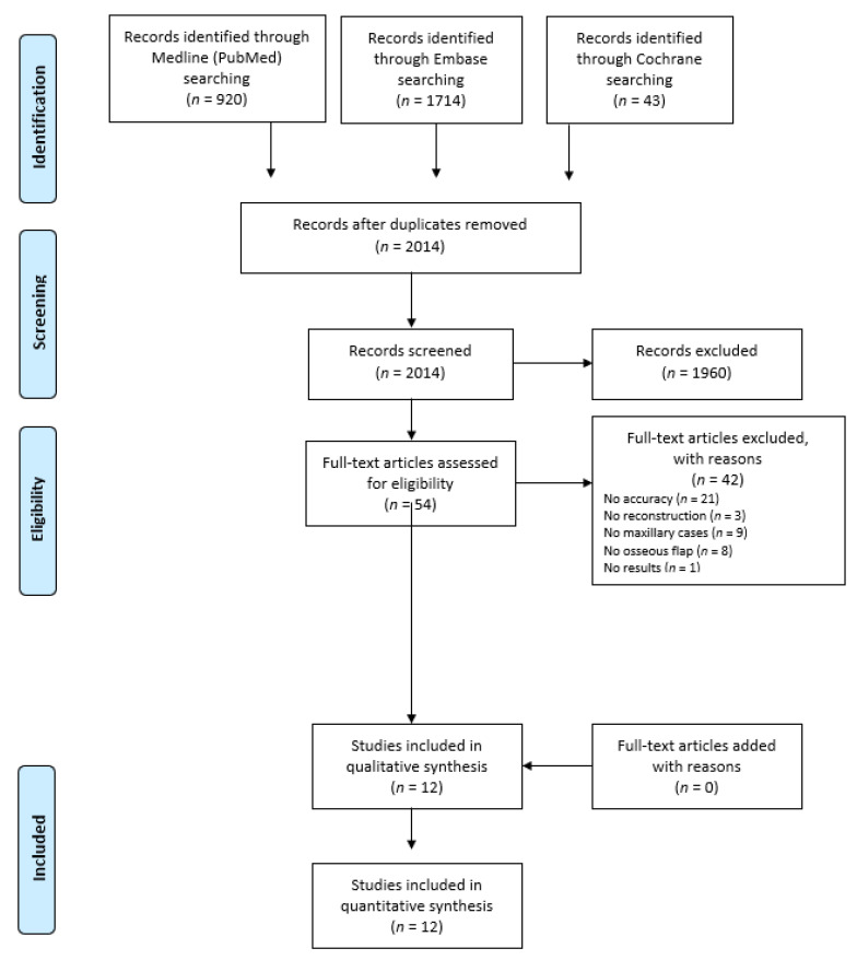
Computer-assisted surgery (CAS) in maxillary reconstruction has proven its value regarding more predictable postoperative results. However, the accuracy evaluation methods differ between studies, and no meta-analysis has been performed yet. A systematic review was performed in the PubMed, Embase, and Cochrane Library databases, using a Patient, Intervention, Comparison and Outcome (PICO) method: (P) patients in need of maxillary reconstruction using free osteocutaneous tissue transfer, (I) reconstructed according to a virtual plan in CAS software, (C) compared to the actual postoperative result, and (O) postoperatively measured by a quantitative accuracy assessment) search strategy, and was reported according to the PRISMA statement. We reviewed all of the studies that quantitatively assessed the accuracy of maxillary reconstructions using CAS. Twelve studies matched the inclusion criteria, reporting 67 maxillary reconstructions. All of the included studies compared postoperative 3D models to preoperative 3D models (revised to the virtual plan). The postoperative accuracy measurements mainly focused on the position of the fibular bony segments. Only approximate comparisons of postoperative accuracy between studies were feasible because of small differences in the postoperative measurement methods; the accuracy of the bony segment positioning ranged between 0.44 mm and 7.8 mm, and between 2.90° and 6.96°. A postoperative evaluation guideline to create uniformity in evaluation methods needs to be considered so as to allow for valid comparisons of postoperative results and to facilitate meta-analyses in the future. With the proper validation of the postoperative results, future research might explore more definitive evidence regarding the management and superiority of CAS in maxillary and midface reconstruction.
Keywords: accuracy; computer-aided design; computer-aided manufacturing; computer-assisted; free tissue flaps; maxillofacial reconstruction; surgery.
Conflict of interest statement
The authors declare no conflict of interest.
Figures
References
-
- Hammond J. Dental care of edentulous patients after resection of maxilla. Br. Dent. J. 1966;120:591–594. - PubMed
-
- Garvey P.B., Chang E.I., Selber J.C., Skoracki R.J., Madewell J.E., Liu J., Yu P., Hanasono M.M. A prospective study of preoperative computed tomographic angiographic mapping of free fibula osteocutaneous flaps for head and neck reconstruction. Plast. Reconstr. Surg. 2012;130:541e–549e. doi: 10.1097/PRS.0b013e318262f115. - DOI - PMC - PubMed
Publication types
LinkOut - more resources
Full Text Sources
Other Literature Sources
