. 2021 Mar 16;11(3):531.
doi: 10.3390/diagnostics11030531.

Dielectric Properties of Ovine Heart at Microwave Frequencies

Affiliations

Dielectric Properties of Ovine Heart at Microwave Frequencies

Niko Ištuk et al. Diagnostics (Basel). .

Abstract

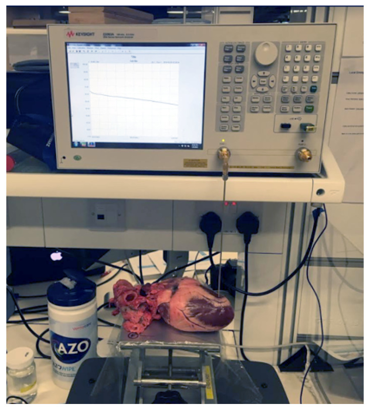
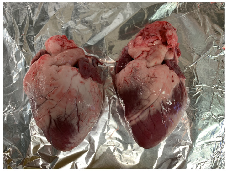
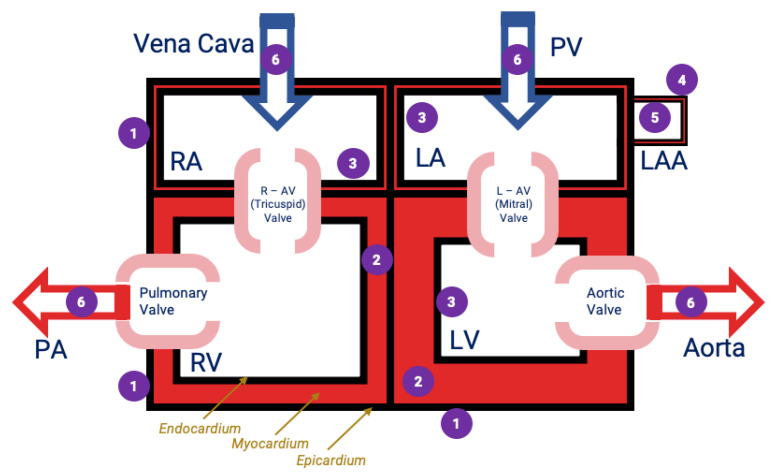
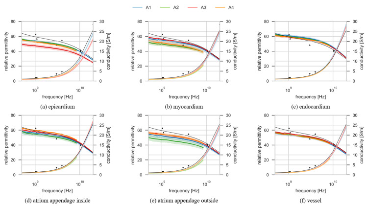
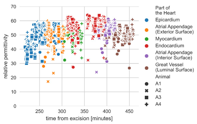
Accurate knowledge of the dielectric properties of biological tissues is important in dosimetry studies and for medical diagnostic, monitoring and therapeutic technologies. In particular, the dielectric properties of the heart are used in numerical simulations of radiofrequency and microwave heart ablation. In one recent study, it was demonstrated that the dielectric properties of different components of the heart can vary considerably, contrary to previous literature that treated the heart as a homogeneous organ with measurements that ignored the anatomical location. Therefore, in this study, we record and report the dielectric properties of the heart as a heterogeneous organ. We measured the dielectric properties at different locations inside and outside of the heart over the 500 MHz to 20 GHz frequency range. Different parts of the heart were identified based on the anatomy of the heart and their function; they include the epicardium, endocardium, myocardium, exterior and interior surfaces of atrial appendage, and the luminal surface of the great vessels. The measured dielectric properties for each part of the heart are reported at both a single frequency (2.4 GHz), which is of interest in microwave medical applications, and as parameters of a broadband Debye model. The results show that in terms of dielectric properties, different parts of the heart should not be considered the same, with more than 25% difference in dielectric properties between some parts. The specific Debye models and single frequency dielectric properties from this study can be used to develop more detailed models of the heart to be used in electromagnetic modeling.

Keywords: ablation; atrial fibrillation; biological tissues; dielectric properties; electromagnetic heating; heart.


Conflict of interest statement

The authors declare no conflict of interest.

Figures

Figure 1
Figure 1
Measurement setup showing VNA2 directly connected to the slim form probe. The sample is brought in contact with the probe by lifting the table while making sure we do not apply excessive probe-sample pressure. The exclusion of the cable from the setup eliminates one source of measurement uncertainty.
Figure 2
Figure 2
Two heart samples from A1 (left) and A2 (right). The hearts are different in size, shape and the amount of pericardial fat covering the epicardium. The pericardial fat extends around the whole heart presenting a challenge when measuring the dielectric properties of the tissues on the exterior surface.
Figure 3
Figure 3
Schematic of measurement locations. n = 17 distinct locations were selected with 15 measurements at each location. These locations were divided into 6 groups as (1) epicardium; (2) endocardium, (3) endocardium, (4) the exterior surface of the atrial appendage, (5) the interior surface of the atrial appendage and (6) luminal surface of the great vessels. These groupings 1–6 are shown in the figure. (RA = right atrium, LA = left atrium, RV = right ventricle, LV = left ventricle, R–AV Valve = right atrioventricular valve, L–AV Valve = left atrioventricular valve, PA = pulmonary artery, PV = pulmonary vein, LAA = left atrial appendage).
Figure 4
Figure 4
Dielectric properties of six parts of the heart (af). Each plot shows relative permittivity and conductivity for one part of the heart, measured on four hearts. Four colors correspond to four hearts: A1 is blue, A2 is green, A3 is red and A4 is orange. Darker lines are relative permittivity and lighter lines are conductivity. The measurement results plotted with the green and the orange lines are measured with the E5063A VNA. The measurement results plotted with the blue and red line are representing the measurements performed with the E8362B VNA. Each line is plotted with the corresponding mean ± 2 standard deviations confidence interval. The thin black lines are the model for the relative permittivity and conductivity of the heart muscle from the literature [10,11,12,51]. The model is based on the data from experimental studies on several different species. The triangle markers are the data points from measurements on human tissues [14].
Figure 5
Figure 5
Single frequency (2.4 GHz) relative permittivity measurements versus time from excision in minutes. Different marker colors represent different parts of the heart. Different marker shapes represent measurements on different hearts. There was no obvious trend observed in changes of dielectric properties measured with respect to time from excision.

Similar articles

Cited by

References

    1. La Gioia A., Porter E., Merunka I., Shahzad A., Salahuddin S., Jones M., O’Halloran M. Open-Ended Coaxial Probe Technique for Dielectric Measurement of Biological Tissues: Challenges and Common Practices. Diagnostics. 2018;8:40. doi: 10.3390/diagnostics8020040. - DOI - PMC - PubMed
    1. Klauenberg B.J., Miklavčič D., editors. Radio Frequency Radiation Dosimetry and Its Relationship to the Biological Effects of Electromagnetic Fields. Springer; Dordrecht, The Netherlands: 2000. - DOI
    1. Chiang J., Hynes K., Brace C.L. Flow-Dependent Vascular Heat Transfer during Microwave Thermal Ablation; Proceedings of the 2012 Annual International Conference of the IEEE Engineering in Medicine and Biology Society; San Diego, CA, USA. 28 August–1 September 2012; pp. 5582–5585. - DOI - PMC - PubMed
    1. Pillai K., Akhter J., Chua T.C., Shehata M., Alzahrani N., Al-Alem I., Morris D.L. Heat Sink Effect on Tumor Ablation Characteristics as Observed in Monopolar Radiofrequency, Bipolar Radiofrequency, and Microwave, Using Ex Vivo Calf Liver Model. Medicine. 2015;94 doi: 10.1097/MD.0000000000000580. - DOI - PMC - PubMed
    1. Chaichanyut M., Tungjitkusolmun S. Microwave Ablation Using Four-Tine Antenna: Effects of Blood Flow Velocity, Vessel Location, and Total Displacement on Porous Hepatic Cancer Tissue. volume 2016. Hindawi; London, UK: 2016. p. E4846738. - DOI - PMC - PubMed

LinkOut - more resources