. 2021 Mar 16;22(6):3030.
doi: 10.3390/ijms22063030.

Apoptotic Effects of Anthocyanins from Vitis coignetiae Pulliat Are Enhanced by Augmented Enhancer of the Rudimentary Homolog (ERH) in Human Gastric Carcinoma MKN28 Cells

Affiliations

Apoptotic Effects of Anthocyanins from Vitis coignetiae Pulliat Are Enhanced by Augmented Enhancer of the Rudimentary Homolog (ERH) in Human Gastric Carcinoma MKN28 Cells

Cheol Park et al. Int J Mol Sci. .

Abstract

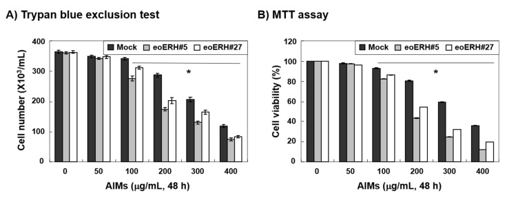
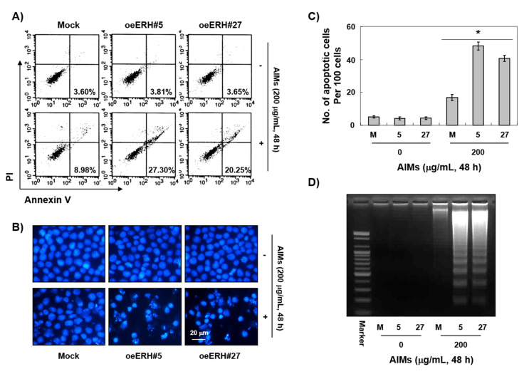
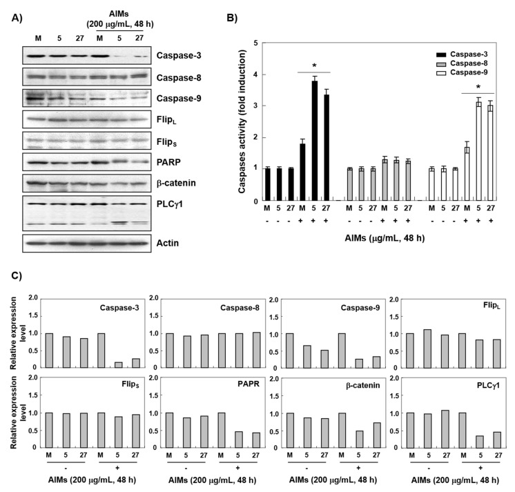
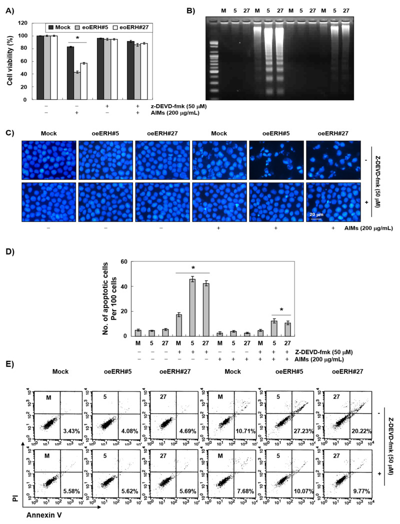
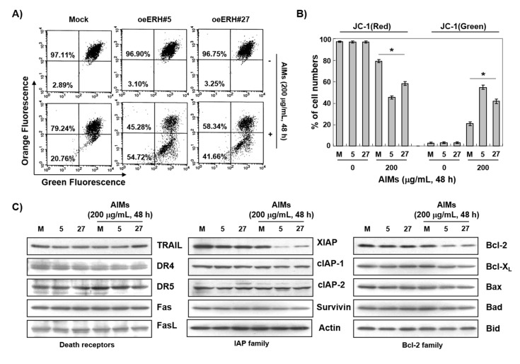
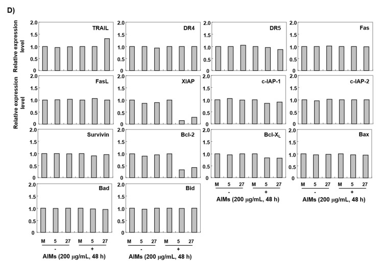
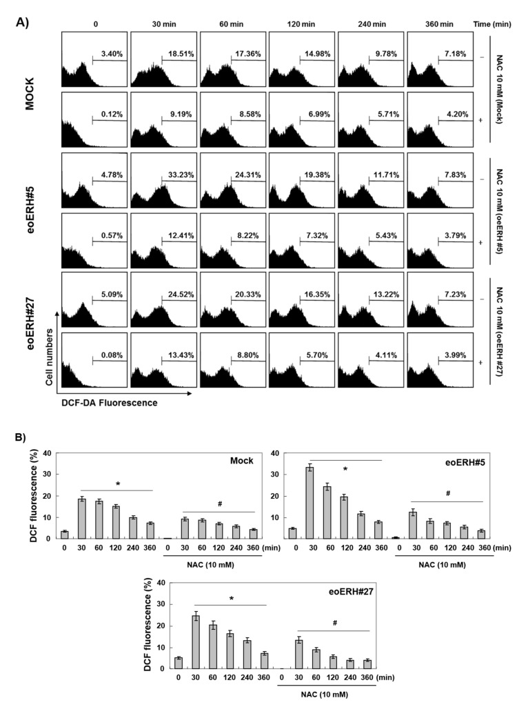
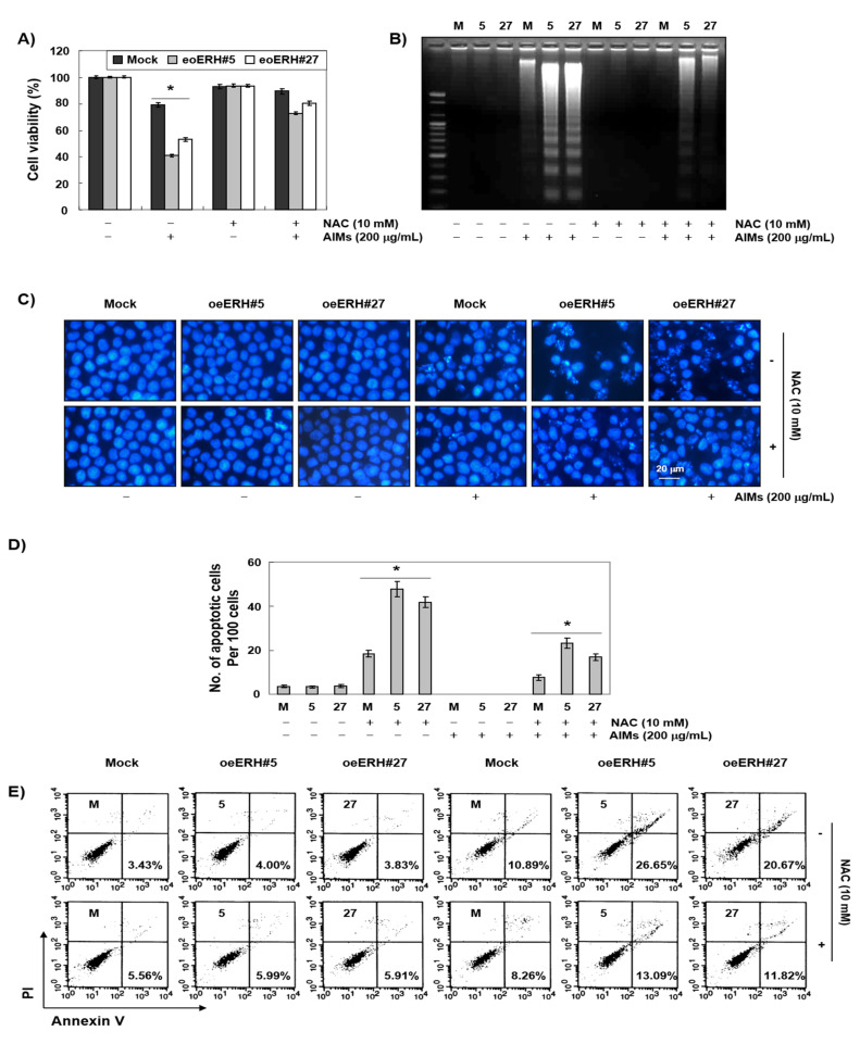
Evidence suggests that augmented expression of a certain gene can influence the efficacy of targeted and conventional chemotherapies. Here, we tested whether the high expression of enhancer of the rudimentary homolog (ERH), which serves as a prognostic factor in some cancers, can influence the efficacy of anthocyanins isolated from fruits of Vitis coignetiae Pulliat, Meoru in Korea (AIMs) on human gastric cancer cells. The anticancer efficacy of AIMs was augmented in ERH-transfected MKN28 cells (E-MKN28 cells). Molecularly, ERH augmented AIM-induced caspase-dependent apoptosis by activating caspase-3 and -9. The ERH-augmented apoptotic effect was related to mitochondrial depolarization and inhibition of antiapoptotic proteins, XIAP, and Bcl-2. In addition, reactive oxygen species (ROS) generation was augmented in AIMs-treated E-MKN28 cells compared to AIMs-treated naïve MKN28 cells. In conclusion, ERH augmented AIM-induced caspase-dependent mitochondrial-related apoptosis in MKN28 cells. A decrease in expression of Bcl-2 and subsequent excessive ROS generation would be the mechanism for ERH-augmented mitochondrial-related apoptosis in AIMs-treated MKN28 cells. A decrease in expression of XIAP would be another mechanism for ERH-augmented caspase-dependent apoptosis in AIMs-treated MKN28 cells.

Keywords: MKN28 human gastric carcinoma cells; Vitis coignetiae Pulliat; anthocyanins; anticancer effects; apoptosis; enhancer of the rudimentary homolog.


Conflict of interest statement

The authors declare no conflict of interest.

Figures

Figure 1
Figure 1
Enhancer of rudimentary homolog (ERH)-augmented antiproliferative effect on AIM-treated MKN28 cells. The cells were seeded at the density of 5 × 10⁴ cells per mL. The two kinds of ERH-transfected MKN28 cells (E-MKN28 cells: eoERH#5 and eoERH#27) and mock-treated MKN28 cells (M-MKN28 cells) were treated with AIMs at indicated concentrations (up to 400 μg/mL). The efficacy of ERH gene on AIM-induced antiproliferative activity was greatest at the concentration of 200 μg/mL of AIMs. The inhibition of cell proliferation was measured by (A) trypan blue exclusion test and (B) MTT assay. The data are shown as means ± SD of three independent experiments. ‘*’ represents significance (* p < 0.05 between AIM-treated E-MKN28 cells and AIM-treated M-MKN28 cells).
Figure 2
Figure 2
ERH-augmented apoptotic effects on AIM-treated MKN28 cells. The cells were seeded at the density of 5 × 10⁴ cells per mL. The two kinds of E-MKN28 cells (eoERH#5 and eoERH#27) and M-MKN28 cells were treated with AIMs at the concentrations of 200 μg/mL for 48 h. ERH increased the apoptotic effects of AIM on MKN28 cells. (A) Annexin V/PI flow cytometry assay, (B) nuclear staining with DAPI solution (Magnification, ×400), and (C) quantitative results for the number of apoptotic cells per 100 cells in total. * p < 0.05 vs. control group. (D) DNA fragmentation test. A ladder pattern of DNA fragmentation indicates internucleosomal cleavage associated with apoptosis. The data are representative of three independent experiments. M, M-MKN28 cells; 5, eoERH#5 E-MKN28 cells; 27, eoERH#27 E-MKN28 cells.
Figure 3
Figure 3
ERH-augmented activation of caspase-3 and -9 in AIM-treated MKN28 cells. The cells were seeded at the density of 5 × 10⁴ cells per mL. The two kinds of E-MKN28 cells (eoERH#5 and eoERH#27) and M-MKN28 cells were treated with AIMs at the concentrations of 200 μg/mL for 48 h. (A) Western blot analysis for procaspase-3, 8, 9, PARP, β-catenin, and PLCγ1 antibodies, (B) caspase activity assay, and (C) densitometry analysis of the data in Western blot analysis by ImageJ software. The values were normalized against β-actin. ERH augmented activation of caspase-3 and -9, in AIMs-treated cells. The data shown as figures are representative of three independent experiments. The data shown in the bar graph are of three independent experiments. ‘*’ represents significance (*p < 0.05 between AIMs-treated E-MKN28 cells and AIMs-treated M-MKN28 cells). M, M-MKN28 cells; 5, eoERH#5 E-MKN28 cells; 27, eoERH#27 E-MKN28 cells.
Figure 4
Figure 4
ERH-augmented caspase-dependent apoptosis in AIM-treated MKN28 cells. The cells were seeded at the density of 5 × 10⁴ cells per mL. The two kinds of E-MKN28 cells (eoERH#5 and eoERH#27) and M-MKN28 cells were treated with AIMs at the concentrations of 200 μg/mL for 48 h. Caspase 3 inhibitor (z-DEVD-fmk) inhibited the ERH-augmented effects on AIM-induced caspase-dependent apoptosis in (A) MTT assay, (B) DNA fragmentation test, (C) nuclear staining with DAPI solution (Magnification, ×400), (D) quantitative results for the number of apoptotic cells per 100 cells in total, and (E) Annexin V/PI flow cytometry assay. The data shown in the bar graph are of three independent experiments. ‘*’ represents significance (*p < 0.05 between groups treated with AIMs alone and treated with AIMs combined with z-DEVD-fmk). The data shown as figures are representative of three independent experiments. M, M-MKN28 cells; 5, eoERH#5 E-MKN28 cells; 27, eoERH#27 E-MKN28 cells.
Figure 5
Figure 5
The mechanisms for ERH-augmented apoptotic effect on AIM-treated cells: mitochondrial depolarization and inhibition of Bcl-2 and XIAP expression. The cells were seeded at the density of 5 × 10⁴ cells per mL. The two kinds of E-MKN28 cells (eoERH#5 and eoERH#27) and M-MKN28 cells were treated with AIMs at the concentrations of 200 μg/mL for 48 h. (A) AIMs induced more loss of MMP (ΔΨm) (mitochondrial depolarization) in E-MKN28 cells than M-MKN28 cells. (B) The data are shown as means ± SD of three independent experiments. ‘*’ represents significance (* p < 0.05 between AIM-treated E-MKN28 cells and AIM-treated M-MKN28 cells). (C) Western blot analysis, which revealed that ERH decreased the expression of antiapoptotic protein XIAP and Bcl-2 in both AIM-untreated and AIM-treated cells. The Western blot analysis data are representative of three independent experiments. (D) Densitometry analysis of the data in Western blot analysis by ImageJ software. The values were normalized against β-actin. (* p < 0.05 between AIM-treated E-MKN28 cells and AIM-treated M-MKN28 cells). M, M-MKN28 cells; 5, eoERH#5 E-MKN28 cells; 27, eoERH#27 E-MKN28 cells.
Figure 5
Figure 5
The mechanisms for ERH-augmented apoptotic effect on AIM-treated cells: mitochondrial depolarization and inhibition of Bcl-2 and XIAP expression. The cells were seeded at the density of 5 × 10⁴ cells per mL. The two kinds of E-MKN28 cells (eoERH#5 and eoERH#27) and M-MKN28 cells were treated with AIMs at the concentrations of 200 μg/mL for 48 h. (A) AIMs induced more loss of MMP (ΔΨm) (mitochondrial depolarization) in E-MKN28 cells than M-MKN28 cells. (B) The data are shown as means ± SD of three independent experiments. ‘*’ represents significance (* p < 0.05 between AIM-treated E-MKN28 cells and AIM-treated M-MKN28 cells). (C) Western blot analysis, which revealed that ERH decreased the expression of antiapoptotic protein XIAP and Bcl-2 in both AIM-untreated and AIM-treated cells. The Western blot analysis data are representative of three independent experiments. (D) Densitometry analysis of the data in Western blot analysis by ImageJ software. The values were normalized against β-actin. (* p < 0.05 between AIM-treated E-MKN28 cells and AIM-treated M-MKN28 cells). M, M-MKN28 cells; 5, eoERH#5 E-MKN28 cells; 27, eoERH#27 E-MKN28 cells.
Figure 6
Figure 6
ERH-augmented ROS generation triggered by AIMs. The cells were seeded at the density of 5 × 10⁴ cells per mL. The two kinds of E-MKN28 cells (eoERH#5 and eoERH#27) and M-MKN28 cells were treated with AIMs at the concentration of 200 μg/mL for 48 h. ROS generation triggered by AIMs is the highest at 30 min after AIMs treatment. At this time, ROS generation is much higher in E-MKN28 cells than M-MKN28 cells. (A) The data shown in the figure are representative of three independent experiments. (B) Summarized flow cytometry data in bar graphs. (*, # p < 0.05 between before and after AIM treatment). M, M-MKN28 cells; 5, eoERH#5 E-MKN28 cells; 27, eoERH#27 E-MKN28 cells.
Figure 7
Figure 7
Another mechanism for ERH-augmented apoptotic effect on AIM-treated cells: excessive ROS generation triggered by AIMs. The cells were seeded at the density of 5 × 10⁴ cells per mL. The two kinds of E-MKN28 cells (eoERH#5 and eoERH#27) and M-MKN28 cells were treated with AIMs at the concentrations of 200 μg/mL for 48 h. With NAC treatment, the ERH-augmented apoptosis was diminished in (A) MTT assay, (B) DNA fragmentation test, (C) nuclear staining with DAPI solution (magnification, ×400), (D) quantitative results for the number of apoptotic cells per 100 cells in total, and (E) Annexin V/PI flow cytometry assay. The data are shown as means ± SD of three independent experiments. ‘*’ represents significance (* p < 0.05 between AIM-treated E-MKN28 cells and AIM-treated M-MKN28 cells). The data shown in figure are representative of three independent experiments. M, M-MKN28 cells; 5, eoERH#5 E-MKN28 cells; 27, eoERH#27 E-MKN28 cells.

Similar articles

Cited by

References

    1. Siegel R.L., Miller K.D., Jemal A. Cancer statistics, 2016. CA Cancer J. Clin. 2016;66:7–30. doi: 10.3322/caac.21332. - DOI - PubMed
    1. Song H.N., Go S.I., Lee W.S., Kim Y., Choi H.J., Lee U.S., Kang M.H., Lee G.W., Kim H.G., Kang J.H., et al. Population-Based Regional Cancer Incidence in Korea: Comparison between Urban and Rural Areas. Cancer Res. Treat. 2016;48:789–797. doi: 10.4143/crt.2015.062. - DOI - PMC - PubMed
    1. Oh C.M., Won Y.J., Jung K.W., Kong H.J., Cho H., Lee J.K., Lee D.H., Lee K.H. Cancer Statistics in Korea: Incidence, Mortality, Survival, and Prevalence in 2013. Cancer Res. Treat. 2016;48:436–450. doi: 10.4143/crt.2016.089. - DOI - PMC - PubMed
    1. Sasako M., Sakuramoto S., Katai H., Kinoshita T., Furukawa H., Yamaguchi T., Nashimoto A., Fujii M., Nakajima T., Ohashi Y. Five-year outcomes of a randomized phase III trial comparing adjuvant chemotherapy with S-1 versus surgery alone in stage II or III gastric cancer. J. Clin. Oncol. 2011;29:4387–4393. doi: 10.1200/JCO.2011.36.5908. - DOI - PubMed
    1. Noh S.H., Park S.R., Yang H.K., Chung H.C., Chung I.J., Kim S.W., Kim H.H., Choi J.H., Kim H.K., Yu W., et al. Adjuvant capecitabine plus oxaliplatin for gastric cancer after D2 gastrectomy (CLASSIC): 5-year follow-up of an open-label, randomised phase 3 trial. Lancet Oncol. 2014;15:1389–1396. doi: 10.1016/S1470-2045(14)70473-5. - DOI - PubMed

MeSH terms