Review
. 2021 Mar 16;10(3):658.
doi: 10.3390/cells10030658.

Synapsins and the Synaptic Vesicle Reserve Pool: Floats or Anchors?

Affiliations
Review

Synapsins and the Synaptic Vesicle Reserve Pool: Floats or Anchors?

Minchuan Zhang et al. Cells. .

Abstract

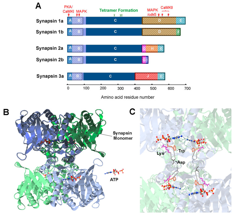
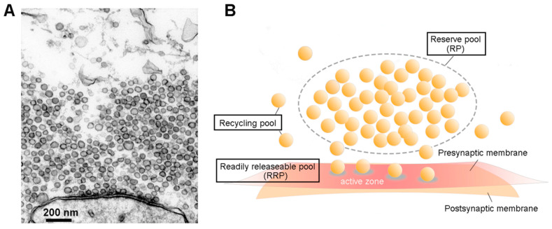
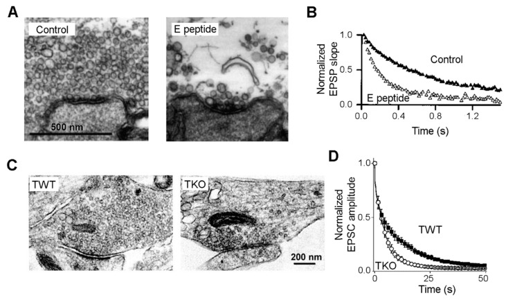
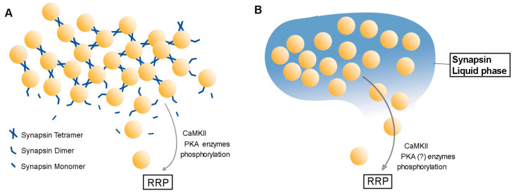
In presynaptic terminals, synaptic vesicles (SVs) are found in a discrete cluster that includes a reserve pool that is mobilized during synaptic activity. Synapsins serve as a key protein for maintaining SVs within this reserve pool, but the mechanism that allows synapsins to do this is unclear. This mechanism is likely to involve synapsins either cross-linking SVs, thereby anchoring SVs to each other, or creating a liquid phase that allows SVs to float within a synapsin droplet. Here, we summarize what is known about the role of synapsins in clustering of SVs and evaluate experimental evidence supporting these two models.

Keywords: neurotransmitter release; presynaptic terminals; synapsins; synaptic vesicle trafficking.


Conflict of interest statement

The authors declare no conflict of interest.

Figures

Figure 2
Figure 2
Synapsin domain structure and tetrameric structure of synapsins. (A). Synapsin isoforms consist of numerous domains, indicated by colors and letters. IDR regions are indicated by shading. Adapted from [41]. (B). Tetrameric structure formed by the C domain of synapsin 2 in the presence of ATP. Two synapsin monomers in a dimer—colored blue or green—are shaded with dark or light colors. A tetramer consists of two dimers. (C). The key residues mediating tetramer formation: aspartate (Asp, black), tryptophan (Trp, orange) and lysine (Lys, magenta); bonds between synapsin dimers are indicated by dashed lines. Structures in (B,C) adapted from data in [42], (accessible at PDB ID: 1i7l), visualized with the NGL viewer [43].
Figure 1
Figure 1
Synaptic vesicles (SVs) and their pools. (A). Electron micrograph of an active zone (AZ) from a squid giant synapse. SVs are the dark, membrane-bound circles that are both attached to the presynaptic plasma membrane and clustered nearby. (B). Classification of SV pools. A reserve pool (RP) occupies the distal volume of the presynaptic terminal, while a readily releasable pool (RRP) consists of vesicles docked at the AZ. The other freely moving SVs are assigned to a recycling pool.
Figure 3
Figure 3
Synapsins and the RP: correlated structural and functional evidence. (A). Injection of a peptide from the E domain of synapsin into squid giant synapses disperses the distal cluster of SVs, while docked SVs remain intact. (B). Synaptic depression, evoked by a 50 Hz train of presynaptic action potentials, is hastened following peptide injection. (A,B adapted from [44]). (C). Comparison of excitatory synapses from synapsin triple-knockout (TKO) and wild-type (TWT) neurons. A distal cluster of SVs is absent in the TKO synapse. (D). Synaptic depression evoked by 10 Hz stimulation is accelerated in TKO neurons, in comparison to TWT neurons. (C,D adapted from [15]).
Figure 4
Figure 4
Reserve pool clustering models. (A). Synaptic vesicle crosslinking model. The vesicles are interlinked by tethers formed by synapsin oligomers. (B). Liquid-liquid phase separation model. Synapsin molecules together with SVs form a distinct liquid phase, trapping the SVs into a cluster.

Similar articles

Cited by

References

    1. Schikorski T., Stevens C.F. Quantitative Ultrastructural Analysis of Hippocampal Excitatory Synapses. J. Neurosci. 1997;17:5858–5867. doi: 10.1523/JNEUROSCI.17-15-05858.1997. - DOI - PMC - PubMed
    1. Alabi A.A., Tsien R.W. Synaptic Vesicle Pools and Dynamics. Cold Spring Harb. Perspect. Biol. 2012;4:a013680. doi: 10.1101/cshperspect.a013680. - DOI - PMC - PubMed
    1. Denker A., Rizzoli S. Synaptic Vesicle Pools: An Update. Front. Synaptic Neurosci. 2010;2:135. doi: 10.3389/fnsyn.2010.00135. - DOI - PMC - PubMed
    1. Rizzoli S.O., Betz W.J. Synaptic vesicle pools. Nat. Rev. Neurosci. 2005;6:57–69. doi: 10.1038/nrn1583. - DOI - PubMed
    1. Rosenmund C., Stevens C.F. Definition of the Readily Releasable Pool of Vesicles at Hippocampal Synapses. Neuron. 1996;16:1197–1207. doi: 10.1016/S0896-6273(00)80146-4. - DOI - PubMed

Publication types

LinkOut - more resources