. 2021 Mar 16;9(3):266.
doi: 10.3390/vaccines9030266.

Escherichia coli Heat-Labile Enterotoxin B Subunit Combined with Ginsenoside Rg1 as an Intranasal Adjuvant Triggers Type I Interferon Signaling Pathway and Enhances Adaptive Immune Responses to an Inactivated PRRSV Vaccine in ICR Mice

Affiliations

Escherichia coli Heat-Labile Enterotoxin B Subunit Combined with Ginsenoside Rg1 as an Intranasal Adjuvant Triggers Type I Interferon Signaling Pathway and Enhances Adaptive Immune Responses to an Inactivated PRRSV Vaccine in ICR Mice

Fei Su et al. Vaccines (Basel). .

Abstract

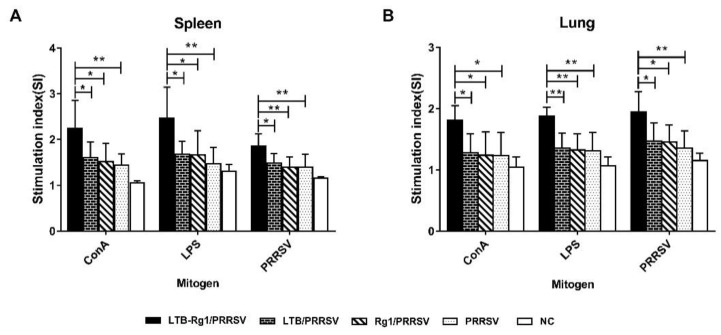
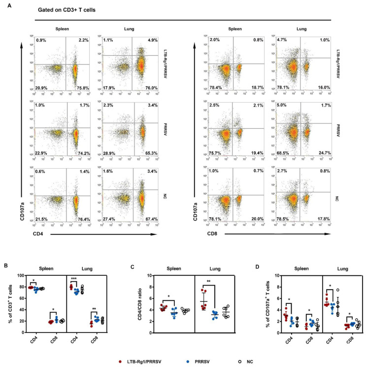
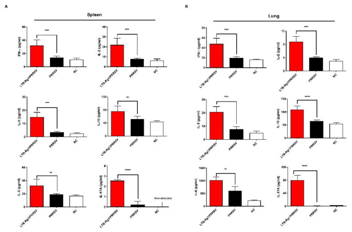
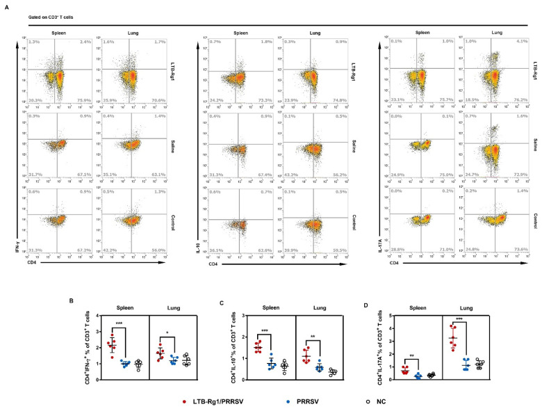
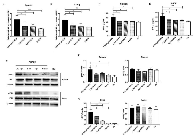
Porcine reproductive and respiratory syndrome virus (PRRSV) is a major pathogen that has threatened the global swine industry for almost 30 years. Because current vaccines do not provide complete protection, exploration of new preventive strategies is urgently needed. Here, we combined a heat-labile enterotoxin B subunit of Escherichia coli (LTB) and ginsenoside Rg1 to form an intranasal adjuvant and evaluated its enhancement of immune responses in mice when added to an inactivated-PRRSV vaccine. The combination adjuvant synergistically elicited higher neutralizing and non-neutralizing (immunoglobulin G and A) antibody responses in the circulatory system and respiratory tract, and enhanced T and B lymphocyte proliferation, CD4+ T-cell priming, and cytotoxic CD4+ T cell activities in mononuclear cells from spleen and lung tissues when compared to the PRRSV vaccine alone, and it resulted in balanced Th1/Th2/Th17 responses. More importantly, we observed that the combination adjuvant also up-regulated type I interferon signaling, which may contribute to improvement in adaptive immune responses. These results highlight the potential value of a combined adjuvant approach for improving the efficacy of vaccination against PRRSV. Further study is required to evaluate the efficacy of this combined adjuvant in swine.

Keywords: LTB; PRRSV; Rg1; intranasal adjuvant; type I interferon.


Conflict of interest statement

The authors declare no conflict of interest.

Figures

Figure 1
Figure 1
Immunization schemes. (A) Vaccination schedule of experiment A. Mice (n = 6/group) were immunized intranasally on days 0, 7, and 21. On day 28, serum and bronchoalveolar lavage (BAL) fluid were collected for antibody assays. (B) Vaccination schedule of experiment B. Mice (n = 48/group) were immunized intranasally on days 0, 7, and 21. On days 7, 14, 21, 28, 35, 42, 49, and 56, 6 mice were taken out from each group and euthanized. Serum and BAL fluid were collected to detect antibody responses. (C) Vaccination schedule of experiment C and D. Mice (n = 18/group in experiment C, n = 12/group in experiment D) were immunized intranasally on days 0, 7, and 21. On day 35, serum and BAL were collected for determination of IgG isotypes and neutralizing antibody titers; spleens and lungs were harvested for detecting IgA-secreting cells, lymphocyte proliferation, cytokine production, T cell differentiation, and type I interferon signaling activities.
Figure 2
Figure 2
B subunit of Escherichia coli heat-labile enterotoxin (LTB-Rg1) increased porcine reproductive and respiratory syndrome virus (PRRSV)-specific antibody responses. After anesthesia with isoflurane, mice (n = 6/group) were intranasally immunized with an inactivated PRRSV vaccine (1 × 10⁵ TCID50) admixed with saline, LTB alone (10–30 µg), Rg1 alone (10–50 µg), or LTB (10–30 µg) combined with Rg1 (10–50 µg) on days 0, 7, and 21. Non-immunized mice served as negative controls. (A) Sera were collected on day 28 for IgG antibody analysis. (B) BAL fluid was collected on day 28 for IgA antibody analysis. Data are expressed as means ± SD. Different letters represent significant difference (p < 0.05).
Figure 3
Figure 3
LTB-Rg1 significantly increased PRRSV-specific serum IgG and respiratory IgA responses. Mice (n = 48/group) were immunized intranasally with PRRSV vaccine admixed with saline, LTB, Rg1, or LTB-Rg1. Non-immunized mice served as negative controls. On days 7, 14, 21, 28, 35, 42, 49, and 56, 6 mice were taken out from each group and euthanized. (A) Sera were collected to detect IgG response. (B) BAL fluid was collected to detect IgA response. Sub-plots show the comparison of LTB-Rg1/PRRSV with PRRSV, LTB/PRRSV, Rg1/PRRSV. Data are expressed as means ± SD. * p < 0.05, ** p < 0.01, *** p < 0.001 versus PRRSV group. # p < 0.05, ## p < 0.01, ### p < 0.001 versus LTB/PRRSV group. p < 0.05, ∆∆ p < 0.01, ∆∆∆ p < 0.001 versus Rg1/PRRSV group.
Figure 4
Figure 4
LTB-Rg1 significantly increased IgG isotypes, IgA-secreting plasma cells, and neutralizing antibody titers. Mice (n = 6/group) were immunized intranasally with PRRSV vaccine admixed with saline, LTB, Rg1, or LTB-Rg1 and euthanized on day 35 post-priming. Non-immunized mice served as negative controls. (A,B) Sera were collected for determination of IgG1 and IgG2a responses. (C) Lung tissues were harvested for detection of IgA-secreting cells by immunohistochemical staining. (D) The integrated optical density (IOD) was analyzed. (E,F) Serum samples and BAL fluid were tested for neutralizing antibody titer. Data are expressed as means ± SD. * p < 0.05, ** p < 0.01, *** p < 0.001. ns, no significant difference.
Figure 5
Figure 5
LTB-Rg1 significantly enhanced lymphocyte proliferative response to ConA, LPS, and PRRSV antigen. Mice (n = 6/group) were immunized intranasally with PRRSV vaccine combined with saline, LTB, Rg1, or LTB-Rg1. Non-immunized mice served as negative controls. Splenocytes and lung mononuclear cells were collected on day 35 and stimulated with ConA, LPS, or PRRSV antigen for 48h. (A) Splenocyte proliferation was determined using CCK-8. (B) The proliferation of lung mononuclear cells was measured. Stimulation index (SI) was calculated. Data are expressed as means ± SD. * p < 0.05, ** p < 0.01.
Figure 6
Figure 6
LTB-Rg1 selectively expanded CD4+ T cell proliferation. Mice (n = 6/group) received PRRSV vaccine without or with LTB-Rg1. Splenocytes and lung mononuclear cells were collected on day 35 post-priming and analyzed by flow cytometry. (A,B) CD4+ and CD8+ T cell subpopulations. (C) CD4+/CD8+ ratio. (D) CD107a+CD4+ and CD107a+CD8+ T cell percentages. Data are expressed as means ± SD. * p < 0.05, ** p < 0.01, *** p < 0.001.
Figure 7
Figure 7
LTB-Rg1 significantly increased cytokine production. Mice (n = 6/group) received PRRSV vaccine without or with LTB-Rg1. Non-immunized animals served as negative controls. On day 35 post-priming, splenocytes and lung cells were prepared and co-cultured with PRRSV antigen for 48 h. (A) Cytokines in the supernatant of splenocytes were tested using ELISA. (B) Cytokines in the supernatant of lung cells were measured. Data are expressed as means ± SD. ** p < 0.01, *** p < 0.001, **** p < 0.0001.
Figure 8
Figure 8
LTB-Rg1 significantly increased cytokine-producing T cells. Mice (n = 6/group) received PRRSV vaccine without or with LTB-Rg1on days 0, 7, and 21. On day 35, splenocytes and lung mononuclear cells were collected and analyzed by flow cytometry. (A,B) Percentages of interferon (IFN)-γ+CD4+ T cells. (A,C) Percentages of IL-10+CD4+ T cells. (A,D) Percentages of IL-17A+CD4+ T cells. Data are expressed as means ± SD. * p < 0.05, ** p < 0.01, *** p < 0.001.
Figure 9
Figure 9
LTB-Rg1 up-regulated type I interferon signaling pathway. Mice (n = 6/group) were immunized intranasally with PRRSV vaccine admixed with either saline, LTB, Rg1, or LTB-Rg1. On day 35, splenocytes and lung mononuclear cells were collected. (A,B) One aliquot each of splenocytes and lung mononuclear cells was re-stimulated with PRRSV antigen for 24 h and tested for interferon regulatory factor (IRF) 3 mRNA expression by qPCR. (C,D) The remaining spleen and lung cells were re-stimulated with PRRSV antigen for 72 h and tested for IFN-α quantification in the supernatant by ELISA. (E) Cell lysis was analyzed for phosphorylation of IRF3 by Western blot. (F,G) The intensity ratios of pIRF3 and IRF3 bands normalized by β-actin in spleen and lung tissues. Data are expressed as means ± SD. * p < 0.05, ** p < 0.01, *** p < 0.001.

Similar articles

Cited by

References

    1. Evans A.B., Loyd H., Dunkelberger J.R., van Tol S., Bolton M.J., Dorman K.S., Dekkers J.C.M., Carpenter S. Antigenic and Biological Characterization of ORF2-6 Variants at Early Times Following PRRSV Infection. Viruses. 2017;9:113. doi: 10.3390/v9050113. - DOI - PMC - PubMed
    1. Nieuwenhuis N., Duinhof T.F., van Nes A. Economic analysis of outbreaks of porcine reproductive and respiratory syndrome virus in nine sow herds. Vet. Rec. 2012;170:225. doi: 10.1136/vr.100101. - DOI - PubMed
    1. Wang H., Du L., Liu F., Wei Z., Gao L., Feng W.H. Highly Pathogenic Porcine Reproductive and Respiratory Syndrome Virus Induces Interleukin-17 Production via Activation of the IRAK1-PI3K-p38MAPK-C/EBPbeta/CREB Pathways. J. Virol. 2019;93 doi: 10.1128/JVI.01100-19. - DOI - PMC - PubMed
    1. Lunney J.K., Fang Y., Ladinig A., Chen N., Li Y., Rowland B., Renukaradhya G.J. Porcine Reproductive and Respiratory Syndrome Virus (PRRSV): Pathogenesis and Interaction with the Immune System. Annu. Rev. Anim. Biosci. 2016;4:129–154. doi: 10.1146/annurev-animal-022114-111025. - DOI - PubMed
    1. Linhares D.C., Cano J.P., Wetzell T., Nerem J., Torremorell M., Dee S.A. Effect of modified-live porcine reproductive and respiratory syndrome virus (PRRSv) vaccine on the shedding of wild-type virus from an infected population of growing pigs. Vaccine. 2012;30:407–413. doi: 10.1016/j.vaccine.2011.10.075. - DOI - PubMed

LinkOut - more resources