

Hepatoprotective Effect of Mixture of Dipropyl Polysulfides in Concanavalin A-Induced Hepatitis
- PMID: 33809904
- PMCID: PMC8004208
- DOI: 10.3390/nu13031022
Hepatoprotective Effect of Mixture of Dipropyl Polysulfides in Concanavalin A-Induced Hepatitis
Abstract
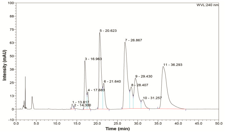
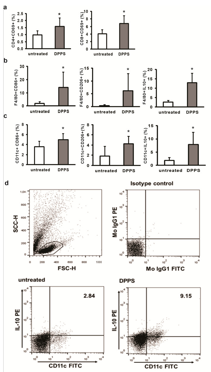
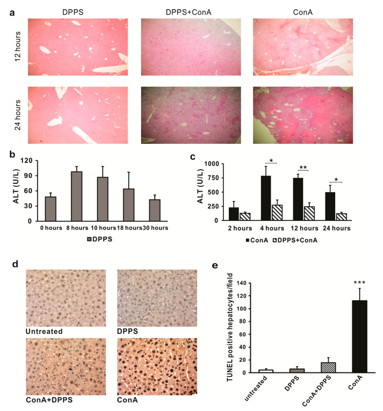
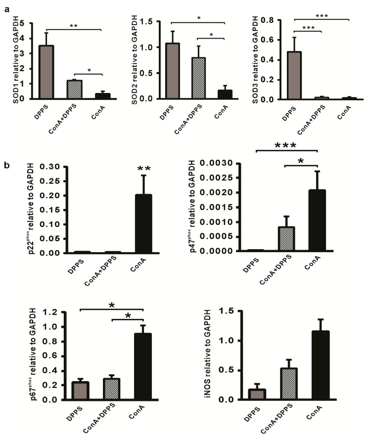
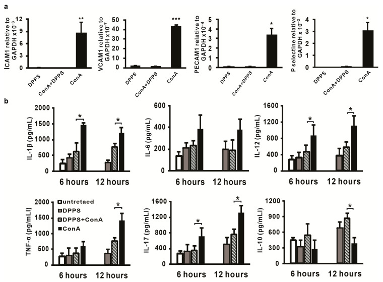
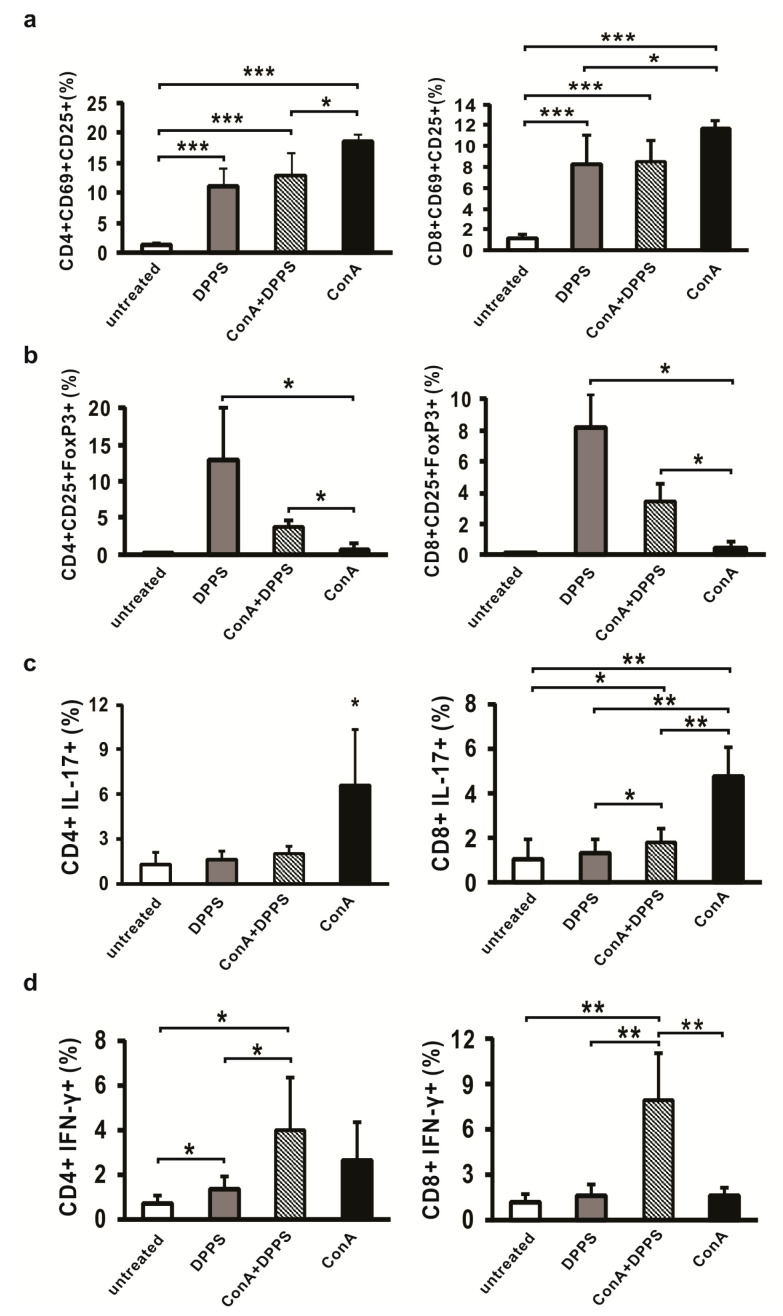
The main biologically active components of plants belonging to the genus Allium, responsible for their biological activities, including anti-inflammatory, antioxidant and immunomodulatory, are organosulfur compounds. The aim of this study was to synthetize the mixture of dipropyl polysulfides (DPPS) and to test their biological activity in acute hepatitis. C57BL/6 mice were administered orally with DPPS 6 h before intravenous injection of Concanavalin A (ConA). Liver inflammation, necrosis and hepatocytes apoptosis were determined by histological analyses. Cytokines in liver tissue were determined by ELISA, expression of adhesive molecules and enzymes by RT PCR, while liver mononuclear cells were analyzed by flow cytometry. DPPS pretreatment significantly attenuated liver inflammation and injury, as evidenced by biochemical and histopathological observations. In DPPS-pretreated mice, messenger RNA levels of adhesion molecules and NADPH oxidase complex were significantly reduced, while the expression of SOD enzymes was enhanced. DPPS pretreatment decreased protein level of inflammatory cytokines and increased percentage of T regulatory cells in the livers of ConA mice. DPPS showed hepatoprotective effects in ConA-induced hepatitis, characterized by attenuation of inflammation and affection of Th17/Treg balance in favor of T regulatory cells and implicating potential therapeutic usage of DPPS mixture in inflammatory liver diseases.
Keywords: ConA hepatitis; anti-inflammatory activity; dipropyl polysulfides; hepatoprotective effects.
Conflict of interest statement
The authors declare no conflict of interest.
Figures
Similar articles
-
15d-PGJ2 alleviates ConA-induced acute liver injury in mice by up-regulating HO-1 and reducing hepatic cell autophagy.Biomed Pharmacother. 2016 May;80:183-192. doi: 10.1016/j.biopha.2016.03.012. Epub 2016 Mar 26. Biomed Pharmacother. 2016. PMID: 27133055
-
Activated farnesoid X receptor attenuates apoptosis and liver injury in autoimmune hepatitis.Mol Med Rep. 2015 Oct;12(4):5821-7. doi: 10.3892/mmr.2015.4159. Epub 2015 Jul 31. Mol Med Rep. 2015. PMID: 26238153 Free PMC article.
-
Total flavonoids from Tetrastigma hemsleyanum ameliorates inflammatory stress in concanavalin A-induced autoimmune hepatitis mice by regulating Treg/Th17 immune homeostasis.Inflammopharmacology. 2019 Dec;27(6):1297-1307. doi: 10.1007/s10787-019-00599-0. Epub 2019 May 23. Inflammopharmacology. 2019. PMID: 31123967
-
Celastrol pretreatment attenuates concanavalin A-induced hepatitis in mice by suppressing interleukin-6/STAT3-interleukin-17 signaling.J Gastroenterol Hepatol. 2023 May;38(5):821-829. doi: 10.1111/jgh.16183. Epub 2023 Mar 26. J Gastroenterol Hepatol. 2023. PMID: 36967570
-
Protective effects of a traditional Chinese herbal formula Jiang-Xian HuGan on Concanavalin A-induced mouse hepatitis via NF-κB and Nrf2 signaling pathways.J Ethnopharmacol. 2018 May 10;217:118-125. doi: 10.1016/j.jep.2018.02.003. Epub 2018 Feb 5. J Ethnopharmacol. 2018. PMID: 29421593
Cited by
-
Concanavalin A promotes angiogenesis and proliferation in endothelial cells through the Akt/ERK/Cyclin D1 axis.Pharm Biol. 2022 Dec;60(1):65-74. doi: 10.1080/13880209.2021.2013259. Pharm Biol. 2022. PMID: 34913414 Free PMC article.
-
Buffering Adaptive Immunity by Hydrogen Sulfide.Cells. 2022 Jan 19;11(3):325. doi: 10.3390/cells11030325. Cells. 2022. PMID: 35159135 Free PMC article. Review.
-
The protective effects of Allium ampeloprasum Subsp Iranicum on cyclophosphamide-induced immunosuppression in NMRI Mice: A promising natural immunomodulator.Avicenna J Phytomed. 2024 Nov-Dec;14(6):734-745. doi: 10.22038/AJP.2024.24595. Avicenna J Phytomed. 2024. PMID: 40259957 Free PMC article.
References
MeSH terms
Substances
LinkOut - more resources
Full Text Sources
Medical
