Review
. 2021 Mar 22;13(6):1441.
doi: 10.3390/cancers13061441.

Key Matrix Remodeling Enzymes: Functions and Targeting in Cancer

Affiliations
Review

Key Matrix Remodeling Enzymes: Functions and Targeting in Cancer

Zoi Piperigkou et al. Cancers (Basel). .

Abstract

Tissue functionality and integrity demand continuous changes in distribution of major components in the extracellular matrices (ECMs) under normal conditions aiming tissue homeostasis. Major matrix degrading proteolytic enzymes are matrix metalloproteinases (MMPs), plasminogen activators, atypical proteases such as intracellular cathepsins and glycolytic enzymes including heparanase and hyaluronidases. Matrix proteases evoke epithelial-to-mesenchymal transition (EMT) and regulate ECM turnover under normal procedures as well as cancer cell phenotype, motility, invasion, autophagy, angiogenesis and exosome formation through vital signaling cascades. ECM remodeling is also achieved by glycolytic enzymes that are essential for cancer cell survival, proliferation and tumor progression. In this article, the types of major matrix remodeling enzymes, their effects in cancer initiation, propagation and progression as well as their pharmacological targeting and ongoing clinical trials are presented and critically discussed.

Keywords: cancer; cathepsins; extracellular matrix; heparanase; hyaluronidases; matrix metalloproteinases; plasminogen activators.


Conflict of interest statement

The authors declare no conflict of interest.

Figures

Figure 1
Figure 1
Prognostic and therapeutic value of MMP14 expression using meta-analysis tools. (AC) Kaplan–Meier survival analysis of relapse-free survival (RFS) probability in different breast cancer patient datasets after diagnosis using gene chip data from GEO, EGA, TCGA databases. MMP14 (MT1-MMP) expression (A) is correlated to better prognosis in ERα-positive (ERα+) breast cancer patients following systemic treatment compared to ERα-negative (ERα-) patients (B) and triple-negative breast cancer patients (C). p value and hazard ratio (HR) value were calculated using a log-rank test [33]. (D) MMP14 gene expression profile of normal mammary tissues, breast invasive carcinoma and metastatic breast tissues, mining RNA-seq and gene chip data from GEO, GTEx, TCGA and TARGET databases, and presented as violin plot. Normal tissue N = 242, Tumor N = 7569, Metastatic N = 82. Abbreviations: EGA, European genome-phenome archive; GEO, gene expression omnibus; GTEx, genotype-tissue expression; TARGET, tumor alterations relevant for genomics-driven therapy; TCGA, the cancer genome atlas.
Figure 2
Figure 2
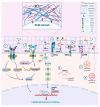
Main functions and signaling cascades mediated by matrix proteolytic enzymes. ➀ TGFβ induces the SMAD-dependent signaling to advance MMP expression. In turn, MMPs in the ECM, as well as uPA may promote the activation of TGFβ to induce cancer-associated EMT. ➁ Matrix remodeling enzymes interact with matrix effectors and affect RTKs activation, thus advancing downstream signaling cascades, like Ras-Raf-MAPK and PI3K/AKT, that lead to increased growth, migration and EMT. ➂ Integrins, major receptors responsible for cell adhesion, contribute to the conversion of pro-MMPs to their active forms. Besides, integrins also facilitate MMP synthesis and regulate their expression levels. Integrin signaling can also regulate the expression and localization of uPA and uPAR, while uPAR interacts with integrins and could act as co-receptor to induce migration and invasion. ➃ Plasmin accelerates syndecan (SDC) ectodomain shedding. ➄ HYALs degrade HA, altering its molecular size and initiating different biological responses, following HA binding to receptors such as CD44. ➅ miRNAs are critical regulators of matrix partners, either by inhibiting or promoting their expression. ➆ HPSE is activated in late endosomes and lysosomes by cathepsin L and stimulates the biogenesis and secretion of exosomes. Additionally, HPSE can be secreted in the ECM under the proper stimuli. ➇ Cathepsins reside in the lysosomes and upon rupture of the lysosomal membrane they are secreted in ECM to exert their actions. On the plasma membrane, they can shed the ectodomains of cell receptors and cleave growth factors and proteins (integrins, E-cadherin etc.). This figure was created using the tools available by BioRender.com, accessed on 2 February 2021.
Figure 3
Figure 3
HPSE gene expression profile of normal mammary tissues, breast invasive carcinoma and metastatic breast tissues mining gene chip data from Gene Expression Omnibus (GEO) repository and presented as boxplot (A) and violin plot (B). Normal tissue N = 242, Tumor N = 7569, Metastatic N = 82 [59].

References

    1. Karamanos N.K., Theocharis A.D., Neill T., Iozzo R.V. Matrix modeling and remodeling: A biological interplay regulating tissue homeostasis and diseases. Matrix Biol. 2019;75:1–11. doi: 10.1016/j.matbio.2018.08.007. - DOI - PMC - PubMed
    1. Iozzo R.V., Gubbiotti M.A. Extracellular matrix: The driving force of mammalian diseases. Matrix Biol. 2018;71:1–9. doi: 10.1016/j.matbio.2018.03.023. - DOI - PMC - PubMed
    1. Borgono C.A., Diamandis E.P. The emerging roles of human tissue kallikreins in cancer. Nat. Rev. Cancer. 2004;4:876–890. doi: 10.1038/nrc1474. - DOI - PubMed
    1. Yang X., Zhang X., Wu R., Huang Q., Jiang Y., Qin J., Yao F., Jin G., Zhang Y. DPPIV promotes endometrial carcinoma cell proliferation, invasion and tumorigenesis. Oncotarget. 2017;8:8679–8692. doi: 10.18632/oncotarget.14412. - DOI - PMC - PubMed
    1. Deryugina E., Carre A., Ardi V., Muramatsu T., Schmidt J., Pham C., Quigley J.P. Neutrophil Elastase Facilitates Tumor Cell Intravasation and Early Metastatic Events. iScience. 2020;23:101799. doi: 10.1016/j.isci.2020.101799. - DOI - PMC - PubMed

LinkOut - more resources