. 2021 Mar 22;22(6):3219.
doi: 10.3390/ijms22063219.

Crystal Structure of a Phospholipase D from the Plant-Associated Bacteria Serratia plymuthica Strain AS9 Reveals a Unique Arrangement of Catalytic Pocket

Affiliations

Crystal Structure of a Phospholipase D from the Plant-Associated Bacteria Serratia plymuthica Strain AS9 Reveals a Unique Arrangement of Catalytic Pocket

Fanghua Wang et al. Int J Mol Sci. .

Abstract

Phospholipases D (PLDs) play important roles in different organisms and in vitro phospholipid modifications, which attract strong interests for investigation. However, the lack of PLD structural information has seriously hampered both the understanding of their structure-function relationships and the structure-based bioengineering of this enzyme. Herein, we presented the crystal structure of a PLD from the plant-associated bacteria Serratia plymuthica strain AS9 (SpPLD) at a resolution of 1.79 Å. Two classical HxKxxxxD (HKD) motifs were found in SpPLD and have shown high structural consistence with several PLDs in the same family. While comparing the structure of SpPLD with the previous resolved PLDs from the same family, several unique conformations on the C-terminus of the HKD motif were demonstrated to participate in the arrangement of the catalytic pocket of SpPLD. In SpPLD, an extented loop conformation between β9 and α9 (aa228-246) was found. Moreover, electrostatic surface potential showed that this loop region in SpPLD was positively charged while the corresponding loops in the two Streptomyces originated PLDs (PDB ID: 1F0I, 2ZE4/2ZE9) were neutral. The shortened loop between α10 and α11 (aa272-275) made the SpPLD unable to form the gate-like structure which existed specically in the two Streptomyces originated PLDs (PDB ID: 1F0I, 2ZE4/2ZE9) and functioned to stabilize the substrates. In contrast, the shortened loop conformation at this corresponding segment was more alike to several nucleases (Nuc, Zuc, mZuc, NucT) within the same family. Moreover, the loop composition between β11 and β12 was also different from the two Streptomyces originated PLDs (PDB ID: 1F0I, 2ZE4/2ZE9), which formed the entrance of the catalytic pocket and were closely related to substrate recognition. So far, SpPLD was the only structurally characterized PLD enzyme from Serratia. The structural information derived here not only helps for the understanding of the biological function of this enzyme in plant protection, but also helps for the understanding of the rational design of the mutant, with potential application in phospholipid modification.

Keywords: Serratia plymuthica strain AS9; bioinformatics; crystal structure; phospholipase D.


Conflict of interest statement

The authors declare no conflict of interest.

Figures

Figure 1
Figure 1
Structure-based amino acid sequence alignment of the mature SpPLD with several other PLDs in the PLDc 2 family. Residues in the conserved HKD motif of the PLD superfamily were indicated with green triangles. For the NucT from Helicobacter pylori (PDB ID: 6EHI), two histidines in the HKD motifs were mutanted to glutamine to obtain enzyme structure in the inactive form.
Figure 2
Figure 2
Phylogenetic analysis of SpPLD with other PLDs. Phylogenetic analysis with neighbor-joining (NJ) was conducted by using MEGA 7.0 software (Hachioji, Tokyo, Japan). Different PLD protein sequeces used for present analysis were shown in the bracket.
Figure 3
Figure 3
Gel filtration chromatography (a) and sodium dodecyl sulphate-polyacrylamide gel electrophoresis (SDS-PAGE) analysis of the purified SpPLD (b).
Figure 4
Figure 4
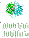
(a) The overall structure of SpPLD. SpPLD was composed of two domains colored with green and light blue, respectively. The active site histidines His116 and H311 were located at the protein active site. The N-terminal domain was composed of 7 α helixs and 7 β sheets and the C-terminal domain was composed by 5 α helixs and 8 β-sheets. (b) Overall topology of SpPLD.
Figure 5
Figure 5
Stereo-view of the superimposition of SpPLD (magenta, PDB ID: 7E0M), Streptomyces sp. PMF PLD (yellow, PDB ID: 1F0I), SaPLD (light blue, PDB ID: 2ZE4), Zuc (orange, PDB ID: 4GEL), NucT(cyans, PDB ID: 6EHI) and Nuc (green, PDB ID: 1BYR).
Figure 6
Figure 6
Two classical HxKxxxxD (HKD) motifs were found in SpPLD and showed high structural consistence with several PLDs within the same family. SpPLD (magentas, PDB ID: 7E0M), Streptomyces sp. PMF PLD (yellow, PDB ID: 1F0I), SaPLD (light blue, PDB ID: 2ZE4), Zuc (orange, PDB ID: 4GEL), NucT(cyans, PDB ID: 6EHI).
Figure 7
Figure 7
(a) Superimpostion and comparision the loop between β9 and α9 with other PLDs. SpPLD (magentas, PDB ID: 7E0M), Streptomyces sp. PMF PLD (yellow, PDB ID: 1F0I), SaPLD (light blue, PDB ID: 2ZE4), Zuc (orange, PDB ID: 4GEL), NucT (cyans, PDB ID: 6EHI), Nuc (green, PDB ID: 1BYR). Two catalytic sites of SpPLD were shown in stick. (b) Sequence alignment of the amino acids that composed the loop between β9 and α9. (c) Electrostatic surface potential profile of the loop between β9 and α9 in SpPLD and the corresponding loop in SaPLD (2ZE4).
Figure 8
Figure 8
(a) Superimpostion and comparision the loop between α10 and α11 with other PLDs. SpPLD (magentas, PDB ID: 7E0M), Streptomyces sp. PMF PLD (yellow, PDB ID: 1F0I), SaPLD (light blue, PDB ID: 2ZE4), Zuc (orange, PDB ID: 4GEL), NucT(cyans, PDB ID: 6EHI), Nuc (green, PDB ID: 1BYR). Two catalytic sites of SpPLD were shown in stick. (b) Sequence alignment of the amino acids that composed the loop between α10 and α11 in SpPLD with other PLDs. (c) Electrostatic surface potential profile of the loop between α10 and α11 of SpPLD.
Figure 9
Figure 9
(a) Superimpostion and comparision the loop between β11 and β12 with other PLDs. SpPLD (magentas, PDB ID: 7E0M), Streptomyces sp. PMF PLD (yellow, PDB ID: 1F0I), SaPLD (light blue, PDB ID: 2ZE4), Zuc (orange, PDB ID: 4GEL), NucT(cyans, PDB ID: 6EHI), Nuc (green, PDB ID: 1BYR). Two catalytic sites of SpPLD were shown in stick. (b) Sequence alignment of the amino acids that composed the loop between β11 and β12 in SpPLD with other PLDs. (c) Electrostatic surface potential profile of the loop between β11 and β12 of SpPLD.
Figure 10
Figure 10
(a) Superimpostion and comparision the C-terminal structure with other PLDs. SpPLD (magentas, PDB ID: 7E0M), Streptomyces sp. PMF PLD (yellow, PDB ID: 1F0I), SaPLD (light blue, PDB ID: 2ZE4), Zuc (orange, PDB ID: 4GEL), NucT(cyans, PDB ID: 6EHI), Nuc (green, PDB ID: 1BYR). Two catalytic sites of SpPLD were shown in stick. (b) Sequence alignment of the amino acids that composed the C-terminal structure in SpPLD with other PLDs.

Similar articles

Cited by

References

    1. Damnjanovi J., Iwasaki Y. Phospholipase D as a catalyst: Application in phospholipid synthesis, molecular structure and protein engineering. J. Biosci. Bioeng. 2013;116:271–280. doi: 10.1016/j.jbiosc.2013.03.008. - DOI - PubMed
    1. Huang T., Lv X., Li J., Shin H.D., Du G., Liu L. Combinatorial fine-tuning of phospholipase D expression by Bacillus subtilis WB600 for the production of phosphatidylserine. J. Microbiol. Biotechnol. 2018;28:2046–2056. doi: 10.4014/jmb.1806.06048. - DOI - PubMed
    1. Hanahan D.J., Chaikoff I.L. A new phospholipide-splitting enzyme specific for the ester linkage between the nitrogenous base and the phosphoric acid grouping. J. Biol. Chem. 1947;169:699–705. doi: 10.1016/S0021-9258(17)30887-6. - DOI - PubMed
    1. Hanahan D.J., Chaikoff I.L. On the nature of the P-containing lipides of cabbage leaves and their relation to a phospholipide-splitting enzyme contained in these leaves. J. Biol. Chem. 1948;172:191–198. doi: 10.1016/S0021-9258(18)35628-X. - DOI - PubMed
    1. Hou H.J., Gong J.S., Dong Y.X., Qin J., Li H., Li H., Lu Z.M., Zhang X.M., Xu Z.H., Shi J.S. Phospholipase D engineering for improving the biocatalytic synthesis of phosphatidylserine. Bioprocess Biosyst. Eng. 2019;42:1185–1194. doi: 10.1007/s00449-019-02116-7. - DOI - PubMed

Substances

Supplementary concepts