. 2021 Mar 22;22(6):3218.
doi: 10.3390/ijms22063218.

Cleavage of the Perlecan-Semaphorin 3A-Plexin A1-Neuropilin-1 (PSPN) Complex by Matrix Metalloproteinase 7/Matrilysin Triggers Prostate Cancer Cell Dyscohesion and Migration

Affiliations

Cleavage of the Perlecan-Semaphorin 3A-Plexin A1-Neuropilin-1 (PSPN) Complex by Matrix Metalloproteinase 7/Matrilysin Triggers Prostate Cancer Cell Dyscohesion and Migration

Tristen V Tellman et al. Int J Mol Sci. .

Abstract

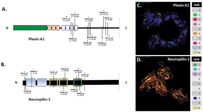
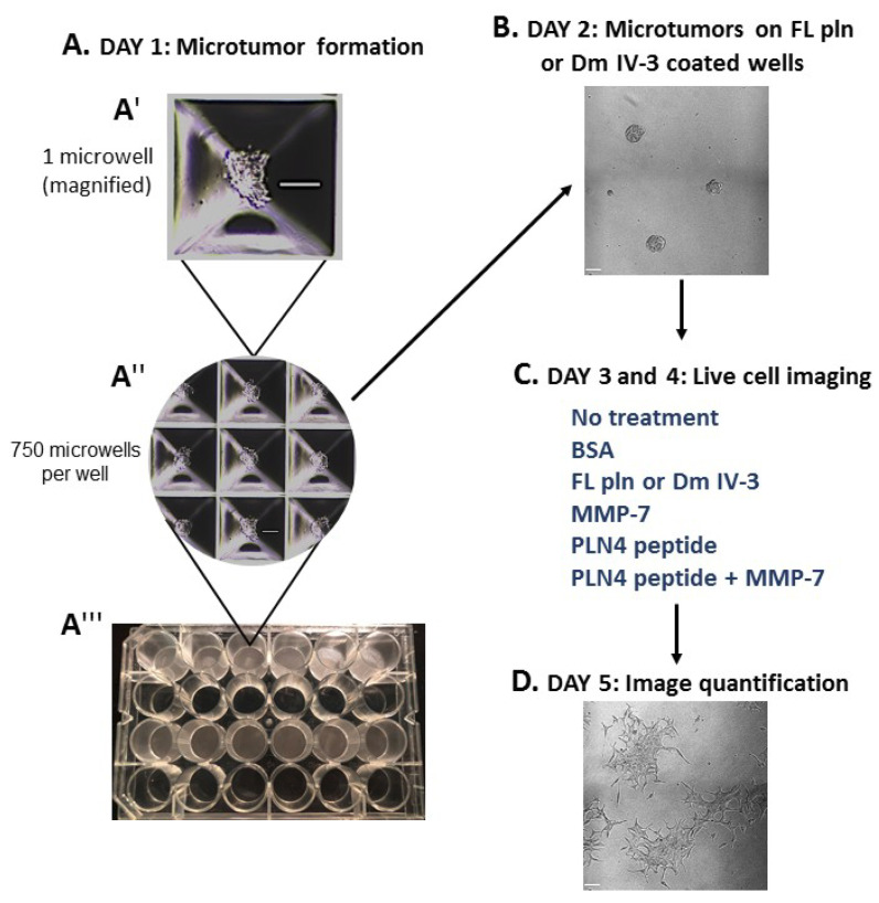
The Perlecan-Semaphorin 3A-Plexin A1-Neuropilin-1 (PSPN) Complex at the cell surface of prostate cancer (PCa) cells influences cell-cell cohesion and dyscohesion. We investigated matrix metalloproteinase-7/matrilysin (MMP-7)'s ability to digest components of the PSPN Complex in bone metastatic PCa cells using in silico analyses and in vitro experiments. Results demonstrated that in addition to the heparan sulfate proteoglycan, perlecan, all components of the PSPN Complex were degraded by MMP-7. To investigate the functional consequences of PSPN Complex cleavage, we developed a preformed microtumor model to examine initiation of cell dispersion after MMP-7 digestion. We found that while perlecan fully decorated with glycosaminoglycan limited dispersion of PCa microtumors, MMP-7 initiated rapid dyscohesion and migration even with perlecan present. Additionally, we found that a bioactive peptide (PLN4) found in perlecan domain IV in a region subject to digestion by MMP-7 further enhanced cell dispersion along with MMP-7. We found that digestion of the PSPN Complex with MMP-7 destabilized cell-cell junctions in microtumors evidenced by loss of co-registration of E-cadherin and F-actin. We conclude that MMP-7 plays a key functional role in PCa cell transition from a cohesive, indolent phenotype to a dyscohesive, migratory phenotype favoring production of circulating tumor cells and metastasis to bone.

Keywords: dyscohesion; matrilysin/MMP-7; microtumors; migration; perlecan/HSPG2; prostate cancer.


Conflict of interest statement

The authors declare no conflict of interest.

Figures

Figure 1
Figure 1
The PSPN Complex forms as a dimer of heterotrimers stabilized by extracellular perlecan. Perlecan/HSPG2 interacts with secreted Sema3A to bind NRP1 (dotted arrow) and plexin A1 to stabilize the PSPN Complex and prevent PCa dyscohesion. Dm IV-3 of perlecan (light green oval) interacts with the Ig module (light green) in the Sema3A dimer. Subsequently, this stabilized dimer interacts with the a1 domain of NRP1 (light blue square), which acts as a bridge between Sema3A and plexin A1. The sema domains of both Sema3A (blue) and plexin A1 (dark green) interact at this a1 NRP1 domain as a dimer of heterotrimers.
Figure 2
Figure 2
In silico predictions demonstrate MMP-7′s potential to cleave plexin A1 and NRP1. In silico digestion of (A) plexin A1 and (B) NRP1 using MMP-7 cleavage sequence information in SitePrediction online software. Schematics of plexin A1 and NRP1 are shown with representative domains and relative location of the top ten predicted cleavage sites. Numbers represent the ranked score for each sequence with the corresponding amino acid sequence, with exact cleavage indicated by the period. PHYRE models of plexin A1 (C) and NRP1 (D) show the predicted ribbon structures with corresponding cleavage sites denoted by color.
Figure 3
Figure 3
In vitro digestions of all PSPN Complex components demonstrate susceptibility to proteolysis by MMP-7. Silver stain of Dm IV-3 incubated with or without MMP-7 (adapted [5]) (A), MMP-7 digestion of Sema3A-Fc (0.75 µg) with or without MMP-7 (0.08 µg) overnight (adapted [4]) (B). Digestion of recombinant plexin A1 (2 µg) (C) and NRP1 (2 µg) (D) overnight with or without MMP-7 (0.2 µg). Black arrows indicate recombinant Dm IV-3, Sema3A-Fc, plexin A1, NRP1 and MMP-7.
Figure 4
Figure 4
Microtumor formation assay. To evaluate the impact of FL pln, MMP-7 and PLN4 peptides on microtumor dispersion, uniformly sized C4-2 microtumors were pre-formed using a 24-well plate microwell system (Kugelmeiers AG. Zürich, Switzerland) (A). One single well of a 24-well plate contains 750 microwells that allowed us to form 750 microtumors of approximately 133 cells per microtumor (A′), (A″). These pre-formed microtumors were transferred to FL pln or Dm IV-3-coated wells for ~24 h (B). Next, microtumors were transferred again to uncoated wells or collagen I-coated wells, and the indicated consecutive treatments in (C) (blue text) were added to the cell culture media. Live-cell imaging (D) stopped at ~48 h and cell dispersion area was quantified (see Figure 5). Scale bars: 100 µm.
Figure 5
Figure 5
Time course analysis of microtumor dispersion under control conditions (no treatment, BSA) or in the presence of FL pln, MMP-7 and/or PLN4 peptide. (A) Representative micrographs showing cell dispersion of C4-2 microtumors with the indicated treatments and time points (scale bar: 100 µm). Yellow dotted lines on the micrographs show the region of interest corresponding to total microtumor dispersion area used to compute area fold change. (B) Evaluation of area fold change. Cell dispersion area is higher in microtumors treated with MMP-7 or MMP-7 in combination with PLN4 peptide compared to microtumors treated with FL pln (see text). Data are the mean ±SEM of three experimental repetitions, except two experimental repetitions for PLN 4 + MMP-7. * p < 0.05, ** p < 0.01. Two-way repeated-measures ANOVA with a post hoc Šídák test was performed. (C) Table shows mean values for fold change in area. Red wireframes demonstrate that the difference in time needed to reach a similar amount of dispersion in the different treatment conditions.
Figure 6
Figure 6
Immunostained microtumors for E-cadherin (red) and F-actin (green) at 24 h. Line scan analyses (see text) were performed for the regions highlighted by the white arrows. Arrow 1: microtumor center, arrow 2: microtumor periphery. Dm IV-3-treated microtumors show increase in co-aligned E-cadherin (red line) and F-actin (green line) at cell–cell contacts (black arrows) whereas microtumors treated with MMP-7 show a decrease in E-cadherin and F-actin co-alignment, indicative of loss of adhesion and initiation of cell dispersion. Line scans were performed on multiple cell–cell boundaries in microtumors under a variety of cohesive and dispersing conditions, then Pearson’s correlation analysis was performed on the same clusters at high magnification (3 clusters/condition). The images shown are representative of what we observed. Scale bars: 30 µm.
Figure 7
Figure 7
Proposed model of the PSPN Complex and E-cadherin/F-actin interaction in cohesive vs. dyscohesive PCa clusters. Model of the dynamic interactions between the PSPN Complex, MMP-7, and E-cadherin/F-actin. Perlecan and Sema3A secreted by the PCa surrounding stroma bind to and silence signaling from the plexin A1-NRP1 proteins. When unproteolyzed, this prevents activation of downstream FAK, AKT, and FOXM1, preventing dyscohesion. At this point, E-cadherin homodimers are intact and the cortical F-actin cytoskeleton is organized near the cell surface. Upon activation of MMP-7, cleavage of the PSPN Complex and extracellular E-cadherin ectodomain occurs, activating integrins, and releasing PCa cells to initiate dyscohesion and migration. Colors and shapes of the PSPN Complex components are described in Figure 1.

Similar articles

Cited by

References

    1. Farach-Carson M.C., Carson D.D. Perlecan a multifunctional extracellular proteoglycan scaffold. Glycobiology. 2007;17:897–905. doi: 10.1093/glycob/cwm043. - DOI - PubMed
    1. Farach-Carson M.C., Warren C.R., Harrington D.A., Carson D.D. Border patrol: Insights into the unique role of perlecan/heparan sulfate proteoglycan 2 at cell and tissue borders. Matrix Biol. 2014;34:64–79. doi: 10.1016/j.matbio.2013.08.004. - DOI - PMC - PubMed
    1. Whitelock J.M., Melrose J., Iozzo R.V. Diverse cell signaling events modulated by Perlecan. Biochemistry. 2008;47:11174–11183. doi: 10.1021/bi8013938. - DOI - PMC - PubMed
    1. Grindel B.J., Martinez J.R., Tellman T.V., Harrington D.A., Zafar H., Nakhleh L., Chung L.W., Farach-Carson M.C. Matrilysin/MMP-7 Cleavage of Perlecan/HSPG2 Complexed with Semaphorin 3A Supports FAK-Mediated Stromal Invasion by Prostate Cancer Cells. Sci. Rep. 2018;8:7262. doi: 10.1038/s41598-018-25435-3. - DOI - PMC - PubMed
    1. Grindel B.J., Martinez J.R., Pennington C.L., Muldoon M., Stave J., Chung L.W., Farach-Carson M.C. Matrilysin/matrix metalloproteinase-7(MMP7) cleavage of perlecan/HSPG2 creates a molecular switch to alter prostate cancer cell behavior. Matrix Biol. 2014;36:64–76. doi: 10.1016/j.matbio.2014.04.005. - DOI - PMC - PubMed

LinkOut - more resources