Review
. 2021 Mar 22;22(6):3224.
doi: 10.3390/ijms22063224.

Mitochondria and Pharmacologic Cardiac Conditioning-At the Heart of Ischemic Injury

Affiliations
Review

Mitochondria and Pharmacologic Cardiac Conditioning-At the Heart of Ischemic Injury

Christopher Lotz et al. Int J Mol Sci. .

Abstract

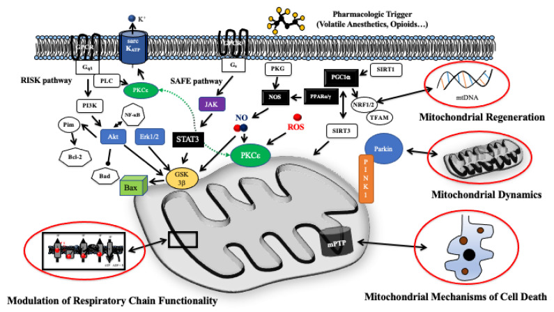
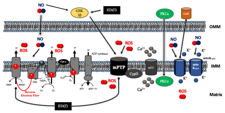
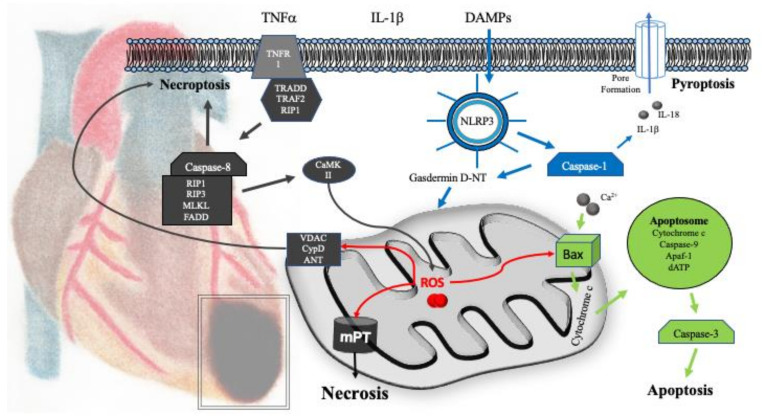
Pharmacologic cardiac conditioning increases the intrinsic resistance against ischemia and reperfusion (I/R) injury. The cardiac conditioning response is mediated via complex signaling networks. These networks have been an intriguing research field for decades, largely advancing our knowledge on cardiac signaling beyond the conditioning response. The centerpieces of this system are the mitochondria, a dynamic organelle, almost acting as a cell within the cell. Mitochondria comprise a plethora of functions at the crossroads of cell death or survival. These include the maintenance of aerobic ATP production and redox signaling, closely entwined with mitochondrial calcium handling and mitochondrial permeability transition. Moreover, mitochondria host pathways of programmed cell death impact the inflammatory response and contain their own mechanisms of fusion and fission (division). These act as quality control mechanisms in cellular ageing, release of pro-apoptotic factors and mitophagy. Furthermore, recently identified mechanisms of mitochondrial regeneration can increase the capacity for oxidative phosphorylation, decrease oxidative stress and might help to beneficially impact myocardial remodeling, as well as invigorate the heart against subsequent ischemic insults. The current review highlights different pathways and unresolved questions surrounding mitochondria in myocardial I/R injury and pharmacological cardiac conditioning.

Keywords: cardioprotection; ischemia/reperfusion injury; preconditioning; volatile anesthetics.


Conflict of interest statement

The authors declare no conflict of interest.

Figures

Figure 1
Figure 1
Schematic depiction of (pre-) mitochondrial signaling in myocardial I/R injury and pharmacologic cardioprotection. Mitochondria host diverse signaling modules at the crossroads of cell survival and cell death. Cytosolic signaling pathways transduce the protective signaling into resulting in modulation of mitochondrial respiratory chain activity and finally the prevention of mitochondria related cell death. Moreover, mitochondrial dynamics and mitochondrial regeneration via transcriptional changes of the mitochondrial proteome modulation contribute to an enhanced intrinsic resistance against I/R injury. Abbreviations: Akt = protein kinase B; Bax = Bcl-2-associated X protein; Bad = Bcl-2-Antagonist of Cell Death; Bcl-2 = B-cell lymphoma-2 protein; Erk1/2 = extracellular regulated kinase 1 and 2; GSK3β = glycogen synthase kinase 3β; Jak = janus-activated kinase; mPTP = mitochondrial permeability transition pore; NO = nitric oxide; NOS = nitric oxide synthase; PI3K = phosphoinositid-3-kinase; PGC1α = peroxisome proliferator-activated receptor gamma coactivator 1-α; Pim = proto-oncogene serine/threonine-protein kinase; PINK1 = PTEN-induced kinase 1; PKCε = protein kinase Cε; PKG = proteinkinase G; PPARα/β = peroxisome proliferator activated receptor α/β; NfκB = nuclear factor κB; NRF1/2 = Nuclear respiratory factor 1/2; RISK = reperfusion injury salvage kinase; sarcKATP = sarcolemmal ATP-dependent potassium channel; SAFE = survival activating factor enhancement; SIRT1/2 = Sirtuin-1/2; STAT3 = signal transducer and activator of transcription 3; TFAM = mitochondrial transcription factor A.
Figure 2
Figure 2
Molecular mechanisms of ischemia/reperfusion injury and cardioprotection acting onto the mitochondria. Diverse intracellular signaling pathways including the RISK (Erk1/2, Akt) and SAFE pathways (Jak) target mitochondrial functions. The centerpiece of cardioprotection is the preservation of mitochondrial respiratory function verhindern massive reactive oxygen species (ROS) production, Ca2+ overload, and subsequent opening of the mitochondrial permeability transition pore (mPT). Abbreviations: Akt = protein kinase B; Cx43 = connexin 43; CypD = cyclophilin D; Erk1/2 = extracellular regulated kinase 1 and 2; GSK3β = glycogen synthase kinase 3β; IMM = inner mitochondrial membrane; Jak = Janus-activated kinase; OMM = outer mitochondrial membrane; NO = nitric oxide; PKCε = protein kinase Cε; RISK = reperfusion injury salvage kinase; SAFE = survival activating factor enhancement; STAT3 = signal transducer and activator of transcription 3.
Figure 3
Figure 3
Mechanisms of cell death after myocardial I/R injury. After hypoxia, myocardial cell death is primarily driven by necrosis subsequent to the loss of mitochondrial function and opening of the mitochondrial permeability transition pore (mPTP). Necrosis also exists as a regulated form of cell death, i.e., necroptosis. Necroptosis is initiated via the activation of death receptors (e.g., tumor necrosis factor receptor 1 (TNFR1). Signaling commences via receptor-interacting protein 1 (RIP1), which mediates the activation of receptor-interacting protein 3 (RIP3) and mixed lineage kinase domain-like (MLKL) alternative activation of calcium-calmodulin-kinase II (CaMKII) by the RIP3/RIP1/MLKL/FADD complex induces mitochondrial dysfunction and membrane permeabilization via cyclophilin D, VDAC, and ANT. Pyroptosis is associated with inflammation subsequent to the ischemic insult and mediated via the NLRP3-inflammasome, caspase-1, and enhanced cytokine production. Mitochondria related apoptosis can also be elicited via mechanisms culminating in the release of cytochrome c and the formation of the apoptosome. Pharmacologic cardioprotection has been shown to act on all forms of cell death by maintaining mitochondrial function, reducing ROS production, as well as direct inhibition of apoptotic signaling or the activation of pro-survival signaling pathways, respectively. Abbreviations: ANT = adenine nucleotide translocator; Apaf-1 = apoptotic protease activating factor 1; Bax = Bcl-2-associated X protein; CaMKII = calcium-calmodulin-kinase II; DAMPs = damage-associated molecular patterns; FADD = Fas-associated protein with death domain; IL-1β = interleukin-1β; IL-18 = interleukin-18; MLKL = mixed lineage kinase domain-like; NLRP3 = NOD-, LRR-, and pyrin domain-containing protein 3; RIP1 = receptor-interacting protein 1; TNFα = tumor necrosis factor α; TNFR1 = tumor necrosis factor receptor 1; TRAF2 = tumor necrosis factor receptor-associated factor 2; TRADD = tumor necrosis factor receptor type 1-associated DEATH domain; VDAC = voltage-dependent anion-selective channel.

References

    1. Yellon D.M., Hausenloy D.J. Myocardial reperfusion injury. N. Engl. J. Med. 2007;357:1121–1135. doi: 10.1056/NEJMra071667. - DOI - PubMed
    1. Murry C.E., Jennings R.B., Reimer K.A. Preconditioning with ischemia: A delay of lethal cell injury in ischemic myocardium. Circulation. 1986;74:1124–1136. doi: 10.1161/01.CIR.74.5.1124. - DOI - PubMed
    1. Tanaka K., Kersten J.R., Riess M.L. Opioid-induced cardioprotection. Curr. Pharm. Des. 2014;20:5696–5705. doi: 10.2174/1381612820666140204120311. - DOI - PMC - PubMed
    1. Kersten J.R. Anesthetic preconditioning: An anesthesiologist’s tale. 1997. Anesthesiology. 2011;114:162–166. doi: 10.1097/ALN.0b013e3181fe4971. - DOI - PubMed
    1. Albrecht M., Zitta K., Bein B., Wennemuth G., Broch O., Renner J., Schuett T., Lauer F., Maahs D., Hummitzsch L., et al. Remote ischemic preconditioning regulates HIF-1alpha levels, apoptosis and inflammation in heart tissue of cardiosurgical patients: A pilot experimental study. Basic Res. Cardiol. 2013;108:314. doi: 10.1007/s00395-012-0314-0. - DOI - PubMed

MeSH terms

Substances

LinkOut - more resources