Review
. 2021 Mar 22;11(3):229.
doi: 10.3390/jpm11030229.

The 35th Anniversary of the Discovery of EPR Effect: A New Wave of Nanomedicines for Tumor-Targeted Drug Delivery-Personal Remarks and Future Prospects

Affiliations
Review

The 35th Anniversary of the Discovery of EPR Effect: A New Wave of Nanomedicines for Tumor-Targeted Drug Delivery-Personal Remarks and Future Prospects

Hiroshi Maeda. J Pers Med. .

Abstract

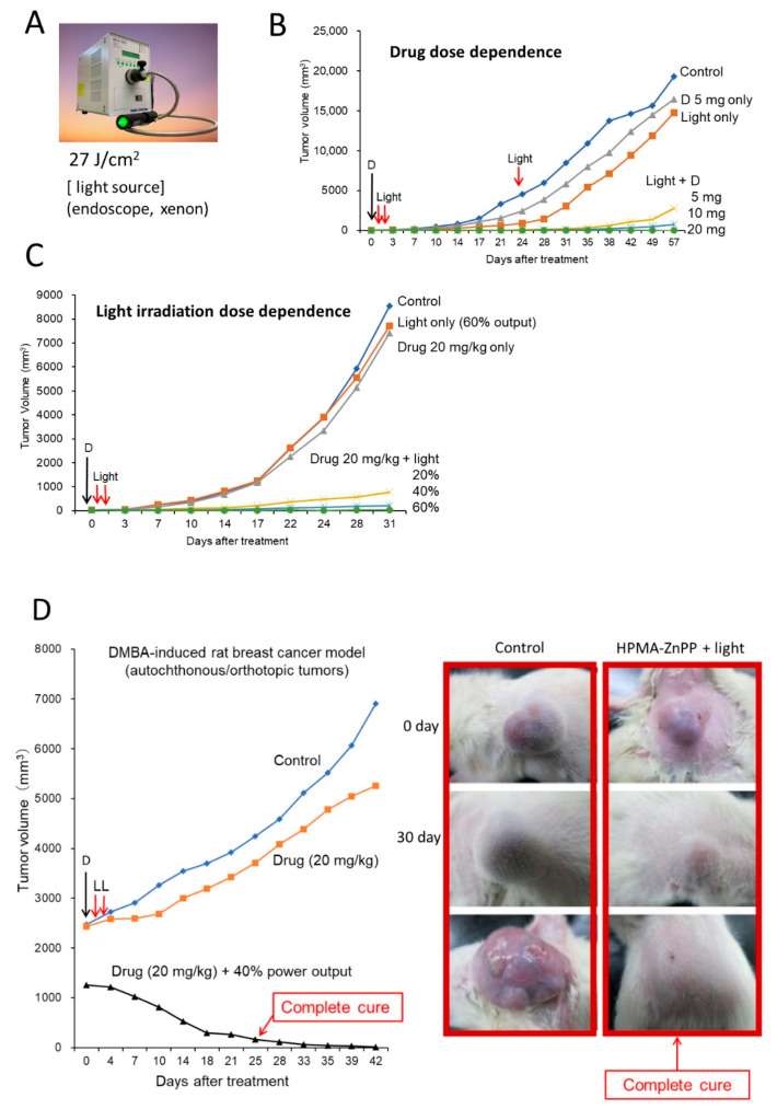
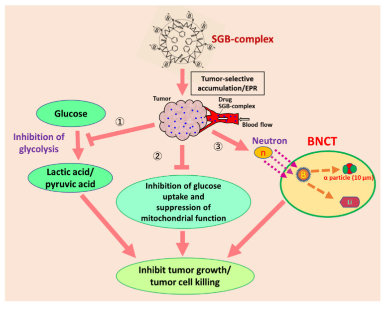
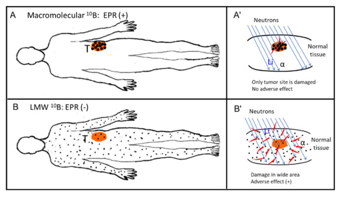
This Special Issue on the enhanced permeability and retention (EPR) effect commemorates the 35th anniversary of its discovery, the original 1986 Matsumura and Maeda finding being published in Cancer Research as a new concept in cancer chemotherapy. My review here describes the history and heterogeneity of the EPR effect, which involves defective tumor blood vessels and blood flow. We reported that restoring obstructed tumor blood flow overcomes impaired drug delivery, leading to improved EPR effects. I also discuss gaps between small animal cancers used in experimental models and large clinical cancers in humans, which usually involve heterogeneous EPR effects, vascular abnormalities in multiple necrotic foci, and tumor emboli. Here, I emphasize arterial infusion of oily formulations of nanodrugs into tumor-feeding arteries, which is the most tumor-selective drug delivery method, with tumor/blood ratios of 100-fold. This method is literally the most personalized medicine because arterial infusions differ for each patient, and drug doses infused depend on tumor size and anatomy in each patient. Future developments in EPR effect-based treatment will range from chemotherapy to photodynamic therapy, boron neutron capture therapy, and therapies for free radical diseases. This review focuses on our own work, which stimulated numerous scientists to perform research in nanotechnology and drug delivery systems, thereby spawning a new cancer treatment era.

Keywords: EPR effect; boron neutron capture therapy; cancer therapy; drug delivery; enhanced permeability and retention effect; nanomedicines; nanotechnology; photodynamic therapy; tumor-selective drug delivery.


Conflict of interest statement

The authors declare no conflict of interest.

Figures

Figure 1
Figure 1
The enhanced permeability and retention (EPR) effect in tumor vasculature. The mechanism of this tumor-selective macromolecular drug targeting depends on various effectors affecting vascular tone, as shown here. Aprotinin is an inhibitor of kallikrein; HOE-140 is a peptide antagonist of kinin. SBTI, soybean trypsin inhibitor; NO, nitric oxide; eNOS, endothelial nitric oxide synthase; iNOS, inducible form of nitric oxide synthase; COXs, cyclooxygenases; PGs, prostaglandins; MMP, metalloproteinase; ONOO, peroxynitrite; O2, superoxide anion radical; MΦ, macrophage; VEGF, vascular endothelial growth factor; VPF, vascular permeability factor; uPA, urokinase plasminogen activator; IL, interleukin; TNF, tumor necrosis factor; B2 receptor, bradykinin B2 receptor (see also Supplementary Figure S1, adapted from ref [23]).
Figure 2
Figure 2
Comparison of the cellular uptake of P-THP—the poly(hydroxypropylmethaacrylamide [HPMA]) conjugate of pirarubicin (THP)—and P-DOX (HPMA polymer-DOX conjugate) by human pancreatic cancer cells (SUIT-2) in vitro. (A) Polymeric P-THP shows a far greater uptake by tumor cells compared with P-DOX: at 30 min, P-THP had a 33.2-fold higher uptake, and its cytotoxicity had greatly increased (see Table at lower left). (B) Penetration of P-DOX, DOX, P-THP, and THP into spheroidal tumor colon cancer (Adapted with permission from ref. [39,40]. 2016 American Chemical Society, 2019 American Chemical Society). Far greater penetration of P-THP into the tumor spheroid (similar to Figure 2, Table) is seen.
Figure 3
Figure 3
Self-assembling PS polymer conjugates of HPMA and ZnPP. (A) Chemical structure of the HPMA-PS polymer conjugate. (B) Polymer-ZnPP in solution. Spontaneous micelles were formed. Quenching occurs in the self-forming micellar form of P-ZnPP, which leads to a lack of fluorescence in the micellar form. When tumor cells take up these micelles, the micelles disintegrate during the traversing lipid bilayer due to its amphiphilic nature. Then, fluorescence becomes positive and singlet oxygen (ROS) are generated in the tumor upon light irradiation (B). ZnPP itself also inhibits heme oxygenase-1 (HO-1) and suppresses tumors (see text for details).
Figure 4
Figure 4
Fluorescence imaging of breast cancer in a rat and of implanted S180 tumor in a mouse, after intravenous injection of P-ZnPP. (A) DMBA (7,12-dimethylbenz[a]anthracene)-induced breast cancer in a rat. Under fluorescent light (left) and under normal light (right). (B) Fluorescent image of nano-PSs: polymeric HPMA-ZnPP (P-HPMA-ZnPP) and free ZnPP. (C) Rhodamine-conjugated bovine albumin (BSA) vs. free rhodamine. Images show no accumulation of LMW free PSs in tumors (B,C). (A, adapted from [58]; B,C, adapted from ref. [4]).
Figure 5
Figure 5
Photodynamic therapy (PDT) with polymeric PSs. (A) View of the light source for the endoscope; a xenon lamp was used. (B) Dose dependence of P-ZnPP dosage, marked D. (C) Dose of light irradiation intensity. The D indicates the time of drug injection of P-ZnPP in B and C. The power of irradiation light (%) is relative to full power output of the endoscope (100%). (D) Results of PDT treatment of DMBA-induced breast cancer in rats. L, light irradiation. D, drug injection. Control received only light. Boxed images at right show growth and suppression of tumor after PDT and P-ZnPP treatment (right) and tumor without treatment (left).
Figure 6
Figure 6
This represents the mode of action of poly(styrene-co-maleic acid) conjugated glucosamine (SGB-complex), which forms complex with boric acid, then forms micelles (~15 nm) and exhibits the EPR effect, about 10 times more boron accumulation in the tumor than other normal tissue [67]. When this SGB-complex is used, it exhibits three different cell killing mechanisms as denoted by “①, ②, and ③” in this figure. By neutron irradiation at right, ③, it elicits the production of α-particles which will kill the tumor cells within 10 micron radius. SGB-complex is rapidly incorporated into the tumor cells and inhibit both glycolysis ① and production of lactic acid; ② it also affects the structural integrity of mitochondria, and its size will shrink and suppress ATP production in the cells (Reprinted with permission from ref. [67]. 2020 Elsevier Ltd.).
Figure 7
Figure 7
Body distribution of boron-containing drugs. (A) Body distribution of a macromolecular 10B compound (e.g., SGB-complex). (B) Distribution of an LMW 10B compound. In (A), boron-containing micelles such as the SGB-complex accumulates predominantly in tumor tissue (T), with the accumulation being about 10 times greater than that of a LMW compound or all other normal tissues in (B). (A’,B’) at right represent enlarged views of the neutron irradiation sites. In (A’), only tumor tissue is damaged: boron micelles (back dots) are evident only in the tumor (T). In (B’), neighboring normal tissue to tumor the boron compound are distributed in most normal tissues such as skin, which will be then be damaged. Red specks around black dots indicate the area of emission of α-particles. (B’) shows that a wide area of tissue is damaged in (B’) (adverse effect).

References

    1. Matsumura Y., Maeda H. A new concept for macromolecular therapeutics in cancer chemotherapy: Mechanism of tumortropic accumulation of proteins and the antitumor agent SMANCS. Cancer Res. 1986;46:6387–6392. - PubMed
    1. Noguchi Y., Wu J., Duncan R., Strohalm J., Ulbrich K., Akaike T., Maeda H. Early phase tumor accumulation of macromolecules: A great difference in clearance rate between tumor and normal tissues. Jpn. J. Cancer Res. 1998;89:307–314. doi: 10.1111/j.1349-7006.1998.tb00563.x. - DOI - PMC - PubMed
    1. Maeda H. Tumor-Selective Delivery of Macromolecular Drugs via the EPR Effect: Background and Future Prospects. Bioconjugate Chem. 2010;21:797–802. doi: 10.1021/bc100070g. - DOI - PubMed
    1. Maeda H. The link between infection and cancer: Tumor vasculature, free radicals, and drug delivery to tumors via the EPR effect. Cancer Sci. 2013;104:779–789. doi: 10.1111/cas.12152. - DOI - PMC - PubMed
    1. Maeda H. Polymer therapeutics and the EPR effect. J. Drug Target. 2017;25:781–785. doi: 10.1080/1061186X.2017.1365878. - DOI - PubMed

LinkOut - more resources