

Prognostic and Functional Significant of Heat Shock Proteins (HSPs) in Breast Cancer Unveiled by Multi-Omics Approaches
- PMID: 33810095
- PMCID: PMC8004706
- DOI: 10.3390/biology10030247
Prognostic and Functional Significant of Heat Shock Proteins (HSPs) in Breast Cancer Unveiled by Multi-Omics Approaches
Abstract
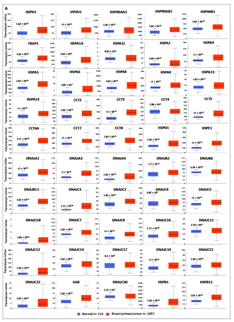
Heat shock proteins (HSPs) are a well-characterized molecular chaperones protein family, classified into six major families, according to their molecular size. A wide range of tumors have been shown to express atypical levels of one or more HSPs, suggesting that they could be used as biomarkers. However, the collective role and the possible coordination of HSP members, as well as the prognostic significance and the functional implications of their deregulated expression in breast cancer (BC) are poorly investigated. Here, we used a systematic multi-omics approach to assess the HSPs expression, the prognostic value, and the underlying mechanisms of tumorigenesis in BC. By using data mining, we showed that several HSPs were deregulated in BC and significantly correlated with a poor or good prognosis. Functional network analysis of HSPs co-expressed genes and miRNAs highlighted their regulatory effects on several biological pathways involved in cancer progression. In particular, these pathways concerned cell cycle and DNA replication for the HSPs co-expressed genes, and miRNAs up-regulated in poor prognosis and Epithelial to Mesenchymal Transition (ETM), as well as receptors-mediated signaling for the HSPs co-expressed genes up-regulated in good prognosis. Furthermore, the proteomic expression of HSPs in a large sample-set of breast cancer tissues revealed much more complexity in their roles in BC and showed that their expression is quite variable among patients and confined into different cellular compartments. In conclusion, integrative analysis of multi-omics data revealed the distinct impact of several HSPs members in BC progression and indicate that collectively they could be useful as biomarkers and therapeutic targets for BC management.
Keywords: HSPs; breast cancer; data mining; expression; miRNAs; prognosis; proteomics.
Conflict of interest statement
The authors declare no conflict of interest.
Figures


Similar articles
-
A multiomics analysis of S100 protein family in breast cancer.Oncotarget. 2018 Jun 26;9(49):29064-29081. doi: 10.18632/oncotarget.25561. eCollection 2018 Jun 26. Oncotarget. 2018. PMID: 30018736 Free PMC article.
-
Comprehensive Pan-Cancer Analysis of Heat Shock Protein 110, 90, 70, and 60 Families.Front Mol Biosci. 2021 Oct 12;8:726244. doi: 10.3389/fmolb.2021.726244. eCollection 2021. Front Mol Biosci. 2021. PMID: 34712697 Free PMC article.
-
Extracellular heat shock proteins and cancer: New perspectives.Transl Oncol. 2021 Feb;14(2):100995. doi: 10.1016/j.tranon.2020.100995. Epub 2020 Dec 15. Transl Oncol. 2021. PMID: 33338880 Free PMC article. Review.
-
Dissecting the functional significance of HSP90AB1 and other heat shock proteins in countering glioblastomas and ependymomas using omics analysis and drug prediction using virtual screening.Neuropeptides. 2023 Dec;102:102383. doi: 10.1016/j.npep.2023.102383. Epub 2023 Sep 14. Neuropeptides. 2023. PMID: 37729687
-
Clinical, Prognostic and Therapeutic Significance of Heat Shock Proteins in Cancer.Curr Drug Targets. 2018;19(13):1478-1490. doi: 10.2174/1389450118666170823121248. Curr Drug Targets. 2018. PMID: 28831912 Review.
Cited by
-
Synergistic Interactions of Cannabidiol with Chemotherapeutic Drugs in MCF7 Cells: Mode of Interaction and Proteomics Analysis of Mechanisms.Int J Mol Sci. 2021 Sep 18;22(18):10103. doi: 10.3390/ijms221810103. Int J Mol Sci. 2021. PMID: 34576262 Free PMC article.
-
Integrated Multi-Omics Investigations of Metalloproteinases in Colon Cancer: Focus on MMP2 and MMP9.Int J Mol Sci. 2021 Nov 17;22(22):12389. doi: 10.3390/ijms222212389. Int J Mol Sci. 2021. PMID: 34830271 Free PMC article.
-
Transcriptomic Analysis of Trachinotus ovatus Under Flow Velocity Stress.Animals (Basel). 2025 Jun 30;15(13):1932. doi: 10.3390/ani15131932. Animals (Basel). 2025. PMID: 40646831 Free PMC article.
-
The Chaperone System in Breast Cancer: Roles and Therapeutic Prospects of the Molecular Chaperones Hsp27, Hsp60, Hsp70, and Hsp90.Int J Mol Sci. 2022 Jul 14;23(14):7792. doi: 10.3390/ijms23147792. Int J Mol Sci. 2022. PMID: 35887137 Free PMC article. Review.
-
Identifying Complex lncRNA/Pseudogene-miRNA-mRNA Crosstalk in Hormone-Dependent Cancers.Biology (Basel). 2021 Oct 9;10(10):1014. doi: 10.3390/biology10101014. Biology (Basel). 2021. PMID: 34681112 Free PMC article.
References
-
- Byler S., Goldgar S., Heerboth S., Leary M., Housman G., Moulton K., Sarkar S. Genetic and epigenetic aspects of breast cancer progression and therapy. Anticancer Res. 2014;34:1071–1077. - PubMed
-
- Gong G., Kwon M.J., Han J., Lee H.J., Lee S.K., Lee J.E., Lee S.H., Park S., Choi J.S., Cho S.Y., et al. A new molecular prognostic score for predicting the risk of distant metastasis in patients with HR+/HER2- early breast cancer. Sci. Rep. 2017;7:45554. doi: 10.1038/srep45554. - DOI - PMC - PubMed
Grants and funding
LinkOut - more resources
Full Text Sources
Other Literature Sources
