Review
. 2021 Mar 22;22(6):3232.
doi: 10.3390/ijms22063232.

Mechanisms of Proteolytic Enzymes and Their Inhibition in QM/MM Studies

Affiliations
Review

Mechanisms of Proteolytic Enzymes and Their Inhibition in QM/MM Studies

Brigitta Elsässer et al. Int J Mol Sci. .

Abstract

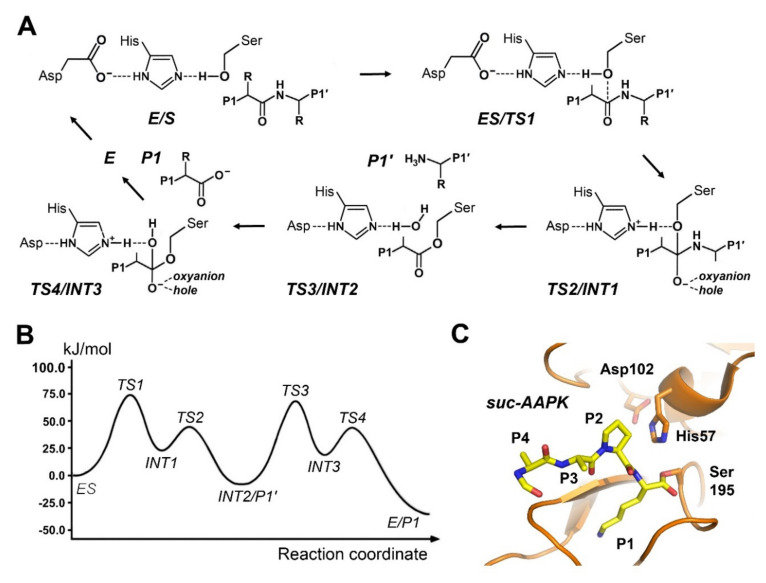
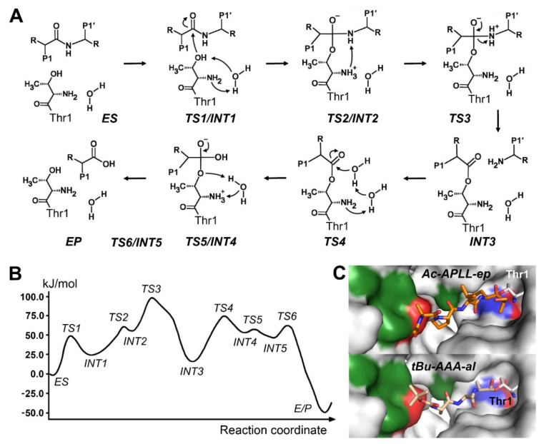
Experimental evidence for enzymatic mechanisms is often scarce, and in many cases inadvertently biased by the employed methods. Thus, apparently contradictory model mechanisms can result in decade long discussions about the correct interpretation of data and the true theory behind it. However, often such opposing views turn out to be special cases of a more comprehensive and superior concept. Molecular dynamics (MD) and the more advanced molecular mechanical and quantum mechanical approach (QM/MM) provide a relatively consistent framework to treat enzymatic mechanisms, in particular, the activity of proteolytic enzymes. In line with this, computational chemistry based on experimental structures came up with studies on all major protease classes in recent years; examples of aspartic, metallo-, cysteine, serine, and threonine protease mechanisms are well founded on corresponding standards. In addition, experimental evidence from enzyme kinetics, structural research, and various other methods supports the described calculated mechanisms. One step beyond is the application of this information to the design of new and powerful inhibitors of disease-related enzymes, such as the HIV protease. In this overview, a few examples demonstrate the high potential of the QM/MM approach for sophisticated pharmaceutical compound design and supporting functions in the analysis of biomolecular structures.

Keywords: enzymes; proteases; qm/mm; quantum chemistry.


Conflict of interest statement

The authors declare no conflict of interest.

Figures

Figure 1
Figure 1
Visualization of the setup for a QM/MM simulation. (A) The molecule of interest, a protease- substrate complex, is centered in a water filled cubic box. Both molecules are depicted with their surface in orange. (B) The MD or MM simulation box shows the protease in ribbon representation (orange), the substrate as ball-and-stick model (green), and the counterions Na+ (purple) and Cl as spheres. (C) Most atoms of the protease in ribbon representation were omitted for clarity, while the QM region with the substrate and the catalytic residues, displayed as ball-and-sticks, is defined by a surrounding, transparent surface. The MM–QM interface requires a special treatment, e.g., by using link atoms.
Figure 2
Figure 2
Mechanism of aspartic proteases. (A) Essentially three major steps takes place, including formation of the Michaelis complex 1. Nucleophilic attack by an activated water molecule with transition state 1 (TS1) and formation of a tetrahedral intermediate (INT). 2. Nitrogen conversion (TS2). 3. Fission of the scissile bond and release of the products with new C- and N-termini (EP). Residue numbering corresponds to pepsin. Two relevant aspartic proteases are shown in Figure 2C. (B) Free energy profile for the pepsin-like protease renin, with an additional reaction step including INT2 and TS3, according to Bras et al. (2012) [33]. (C) Left panel: Active site of pepsin with a phosphonate inhibitor, mimicking TS2 (PDB 1QRP). White areas are polar, green areas are hydrophobic. Right panel: The dimeric HIV protease has one catalytic Asp25 (red spheres) per monomer (PDB 4HVP). A peptidic inhibitor (purple sticks) is bound to the active site as ES analog.
Figure 3
Figure 3
(A) Mechanism of metalloproteases. 1. Formation of a Michaelis complex (ES) and binding of the carbonyl O of the P1 residue to the catalytic Zn2+, which functions as oxyanion hole. 2. Nucleophilic attack by an activated water molecule, i.e., OH (TS1), formation of a tetrahedral intermediate and transfer of an H+ to the amide NH of the scissile bond (INT) 3. Cleavage of the scissile bond (TS2) and product release (E/P). Especially step 2 can be further subdivided into more steps. Structural examples with functional relevance are shown in Figure 3C. (B) The relatively simple free energy profile for MMP3 follows Pelmenshikov and Siegbahn (2002) [87]. (C) Left panel: MMP1 in complex with the natural substrate collagen (PDB 966C) corresponds to the ES complex. Right panel: MMP1 active site with a Zn2+ bound hydroxamate inhibitor (PDB 4AUO), which partially resembles the intermediate (INT).
Figure 4
Figure 4
Mechanism of cysteine proteases. (A) As in the prototypic papain the catalytic residues of the dyad are a Cys and a His. Essentially two major reaction steps take place, namely the acylation and the deacylation, while several sub steps are involved, according to Wei et al. (2013) [99]. 1. Nucleophilic attack by the negatively charged Sγ atom on the carbonyl C of the P1 residue (TS1) and formation of tetrahedral intermediate (INT1). The oxyanion hole stabilizes the negative charge at the carbonyl O atom. 2. Upon protonation of the amide NH group the scissile bond breaks and the P1′ product leaves with a new N-terminus (TS2). 3. The acyl intermediate (INT2) is attacked by the nucleophilic catalytic water (TS3) and forms the second tetrahedral intermediate (INT3). 4. Release of the P1 product with a new carboxy terminus (TS4/INT4 and E/P1). (B) Free energy profile of the above described reaction. (C) The coronavirus SARS-Cov-2 main protease (MPro) is depicted as free enzyme dimer (PDB 6Y2E) on the left and with a covalent ketoamide inhibitor (PDB 6Y2G) as TS2 analog on the right. Some residues of the protease were omitted for clarity.
Figure 5
Figure 5
Mechanism of serine proteases. (A) The basic mechanism resembles the one of cysteine proteases. In a catalytic triad the acidic Asp is required to stabilize the positively charged His, which enhances the nucleophilicity of the Ser Oγ atom. 1. Formation of the Michaelis complex (ES). 2. Nucleophilic attack by the negatively polarized Oγ atom to the carbonyl C of the P1 residue (TS1), resulting in the tetrahedral intermediate with a negative charge at the carbonyl O (INT1), which is bound to oxyanion hole. 3. Protonation of the amide NH group breaks the scissile bond (TS2), with release of the of the P1′ product. 4. The acyl intermediate (INT2) is attacked by the catalytic water (TS3) and forms the second tetrahedral intermediate (INT3). 5. Release of the P1 product (TS4/E/P1) with a new C-terminus. (B) Free energy profile of the reaction in trypsin. (C) Trypsin in complex with a succinyl-Ala-Ala-Pro-Lys inhibitor (PDB 2AGG), which forms a true acyl intermediate, corresponding to INT2.
Figure 6
Figure 6
Mechanistic details of threonine protease catalysis. (A) The six step mechanism according to Wei et al., 2013 [175]. (B) Free energy profile of the six-step reaction. (C) Inhibitor structures of the yeast 20S proteasome. Upper panel: The epoxide Ac-Ala-Pro-Leu-Leu-ep covalently bound to the Thr1 Oγ atom of the β5 subunit corresponds to the tetrahedral intermediate INT2. The αN-C bond is not shown for clarity in the upper panel (PDB 4QBY). Lower panel: The tBu-Ala-Ala-Ala-aldehyde bound to Thr1 represents the acyl intermediate INT3 (PDB 4Y8L).

References

    1. Bond J.S. Proteases: History, discovery, and roles in health and disease. J. Biol. Chem. 2019;294:1643–1651. doi: 10.1074/jbc.TM118.004156. - DOI - PMC - PubMed
    1. Levene P.A. The Cleavage Products of Proteoses. J. Biol. Chem. 1905;1:45–58. doi: 10.1016/S0021-9258(17)46095-9. - DOI
    1. Robertson T.B. Note on the Synthesis of A Protein through the Action of Pepsin. J. Biol. Chem. 1907;3:95–99. doi: 10.1016/S0021-9258(17)45998-9. - DOI
    1. Northrop J.H. Crystalline Pepsin. Science. 1929;69:580. doi: 10.1126/science.69.1796.580. - DOI - PubMed
    1. Bernal J.D., Crowfoot D. X-Ray Photographs of Crystalline Pepsin. Nature. 1934;133:794–795. doi: 10.1038/133794b0. - DOI

LinkOut - more resources