. 2021 Mar 22;22(6):3233.
doi: 10.3390/ijms22063233.

Murine Esophagus Expresses Glial-Derived Central Nervous System Antigens

Affiliations

Murine Esophagus Expresses Glial-Derived Central Nervous System Antigens

Christopher Kapitza et al. Int J Mol Sci. .

Abstract

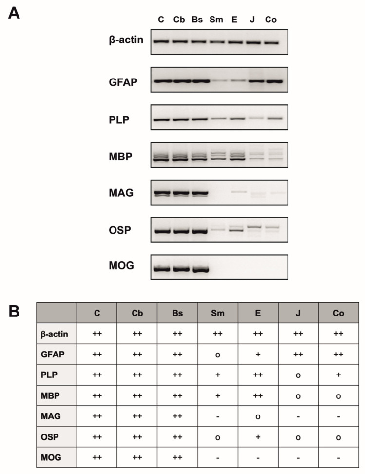
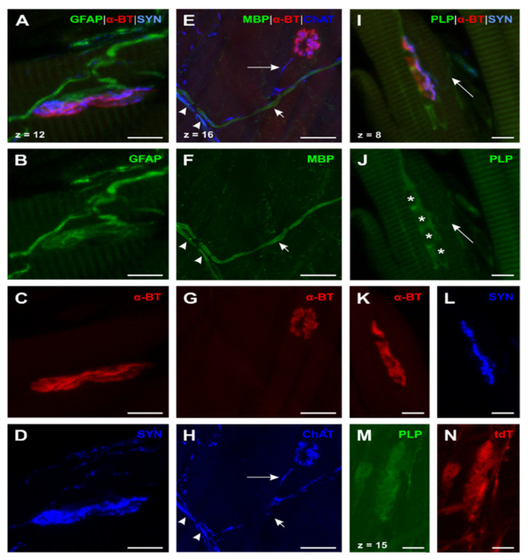
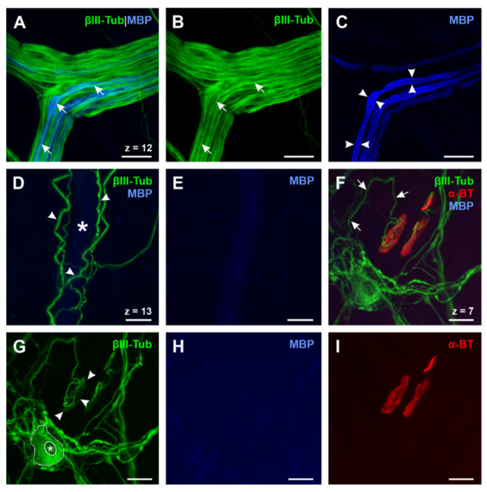
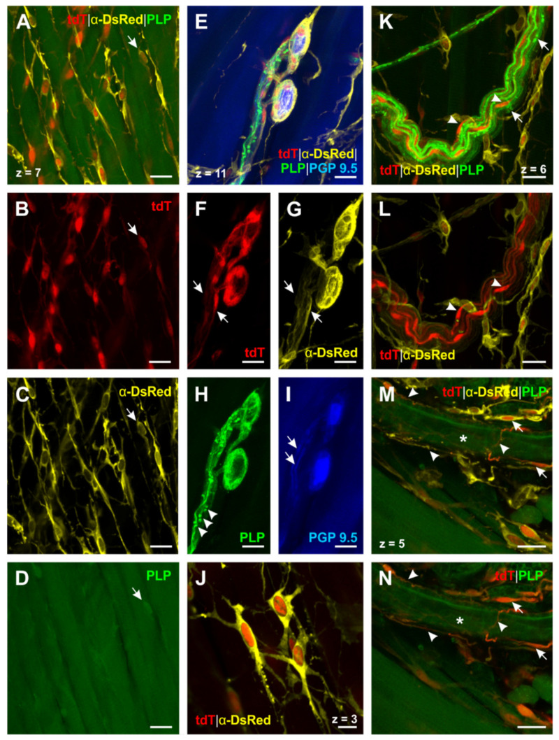
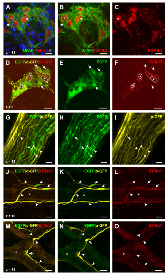
Multiple sclerosis (MS) has been considered to specifically affect the central nervous system (CNS) for a long time. As autonomic dysfunction including dysphagia can occur as accompanying phenomena in patients, the enteric nervous system has been attracting increasing attention over the past years. The aim of this study was to identify glial and myelin markers as potential target structures for autoimmune processes in the esophagus. RT-PCR analysis revealed glial fibrillary acidic protein (GFAP), proteolipid protein (PLP), and myelin basic protein (MBP) expression, but an absence of myelin oligodendrocyte glycoprotein (MOG) in the murine esophagus. Selected immunohistochemistry for GFAP, PLP, and MBP including transgenic mice with cell-type specific expression of PLP and GFAP supported these results by detection of (1) GFAP, PLP, and MBP in Schwann cells in skeletal muscle and esophagus; (2) GFAP, PLP, but no MBP in perisynaptic Schwann cells of skeletal and esophageal motor endplates; (3) GFAP and PLP, but no MBP in glial cells surrounding esophageal myenteric neurons; and (4) PLP, but no GFAP and MBP in enteric glial cells forming a network in the esophagus. Our results pave the way for further investigations regarding the involvement of esophageal glial cells in the pathogenesis of dysphagia in MS.

Keywords: autoantibodies; dysphagia; enteric glia; enteric nervous system; esophagus; glial fibrillary acidic protein; motor endplate; multiple sclerosis; myelin basic protein; proteolipid protein.


Conflict of interest statement

The authors declare no conflict of interests.

Figures

Figure 1
Figure 1
RT-PCR analysis for β-actin, glial fibrillary acidic protein (GFAP), proteolipid protein (PLP), myelin basic protein (MBP), myelin associated glycoprotein (MAG), oligodendrocyte-specific protein (OSP) and myelin oligodendrocyte glycoprotein (MOG) of n = 6 mice. (A) Overview of different expression levels of the examined markers with β-actin used as housekeeping gene, shown by one representative mouse. (B) Table summarizing the PCR results of six mice and showing the expression profile of the different tissues investigated with + indicating a “high expression”, + indicating a “medium–low expression”, o indicating a “very low expression” and–indicating no expression. Since results from individual mice did not differ, data were pooled. C: Cerebrum; Cb: Cerebellum; Bs: Brain stem; Sm: Skeletal muscle (M. tibialis anterior); E: Esophagus; J: Jejunum; Co: Colon ascendens; GFAP: Glial fibrillary acidic protein; PLP: Proteolipid protein; MBP: Myelin basic protein; MAG: Myelin-associated glycoprotein; OSP: Oligodendrocyte-specific protein; MOG: Myelin oligodendrocyte glycoprotein.
Figure 2
Figure 2
Expression of GFAP, MBP, and PLP in neuromuscular junction (NMJ) of the esophagus (A)–(D): Triple staining for GFAP, α-bungarotoxin (α-BT), and synaptophysin (SYN) (N° ③, Table 1) showed a similar distribution as the skeletal muscle with the exception that efferences were unmyelinated and had a smaller caliber. (E)–(H): Quadruple staining for MBP, α-BT, cholin acetyltransferase (ChAT), and Hoechst (Hoechst not shown; N° ①, Table 2). ChAT+-efferences which contact the motor endplate always lack myelin ((E) and (H), long arrow). Two further types of fibers could be detected: (1) Myelinated ChAT+-efferences ((E) and (F), arrowheads) and (2) myelinated ChAT -fibers ((E) and (F), short arrow)—the latter can be brought in line with esophageal afferences. (I) and (L): Quadruple staining for PLP, α-BT, SYN, and Hoechst (Hoechst not shown; N° ⑤; Table 1). In contrast to the skeletal muscle, PLP was present in all investigated NMJs as grouped PLP+ -glial cells around the endplate indicate (I) and (J). Nuclei of these PLP+ -PSCs are marked by asterisks (J); confirmed by Hoechst nuclear staining (not shown)). Contrary to the distribution of MBP, PLP could also be found throughout the Plexus myentericus as PLP+-efferences show ((I) and (J), long arrow). (M) and (N): Triple staining for Discosoma sp. red fluorescent protein (DsRed1), PLP, and protein gene product 9.5 (PGP 9.5) in PLP-CreERT2 x tdTomato (tdT) mice (anti-DsRed and PGP 9.5 not shown; N° ②, Table 2). The faint signal of PLP in the endplate region was confirmed by tdT expression as both PLP and tdT show the same distribution (M) and (N). α-BT: α-bungarotoxin; ChAT: Cholin acetyltransferase; DsRed: Discosoma sp. red fluorescent protein; GFAP: Glial fibrillary acidic protein; MBP: Myelin basic protein; PGP 9.5: Protein gene product 9.5; PLP: Proteolipid protein; SYN: Synaptophysin; tdT: tdTomato. Z-step = 1 µm (A)–(D); (I)–(N) and 0.8 µm (E)–(H); scale bars 10 µm (A)–(D), (I)–(N), 30 µm (E)–(H).
Figure 3
Figure 3
Expression of βIII-Tubulin, MBP, and α-BT in the esophagus (A)–(E): Triple whole mount staining of βIII-tubulin, MBP, and α-BT (α-BT not shown, N° ⑤, Table 2). Axons ((A) and (B), short arrows) of peripheral vagal nerve branches are only partially myelinated (C, arrowheads), while gracile blood vessel-related nerve fibers, which are tightly wrapped around the outer vessel wall, appear βIII-tubulin-positive (D, arrowheads) but always lack myelin since no MBP signal can be detected (E). The lumen of the blood vessel is marked by the asterisk (D). (FI): Triple whole mount staining of βIII-tubulin, MBP and α-BT (N° ⑤, Table 2) reveals that endplate contacting efferent axons (F, short arrows) are always unmyelinated for a long distance (H) and form a framework in the presynaptic region of the NMJ (G, arrowheads). These findings confirm the results of the ChAT staining protocol (cf. Figure 2E–H). Moreover, βIII-tubulin can also be found in enteric neurons (G, dotted line; nucleus marked by asterisk) of the myenteric plexus and therefore proves to be a suitable neuro-axonal marker for the evaluation of the ENS in the esophagus. ChAT: Cholin acetyltransferase; MBP: Myelin basic protein; Z-step = 1 µm; scale bars 20 µm.
Figure 4
Figure 4
PLP+ -glial cells in the esophagus identified by triple staining for DsRed, PLP and PGP 9.5 in PLP-CreERT2 x tdT mice (N° ②, Table 2). (A)–(D) (PGP 9.5 not shown): PLP+ -glial cells form a meshwork throughout the esophagus. Some of these cells, located in the tunica muscularis, are arranged in parallel with the muscle fibers. While only a few of these cells can be detected by PLP antibody staining ((AD), short arrow), transgenic PLP-CreERT2 x tdT mice reveal their distribution (cf. AC vs. D). (EI): PLP+ -glial cells interact very closely with enteric neurons as the processes of the EGCs are woven around the neurons (EH). Interconnecting strands of enteric neurons (I, short arrows) are accompanied by PLP+ -glial cells (F and G, short arrows). PLP antibody staining indicates the contact zone of these cells (H, arrowheads). (J) (PGP 9.5 and PLP not shown): Close-up of PLP+ -EGCs; these cells are interconnected by their fine, filiform processes and therefore form a network of glial cells. (KL) (PGP 9.5 not shown): PLP+ -myelin sheaths of a vagal nerve fiber bundle in the tunica adventitia can be identified by the PLP antibody (K, nodes of Ranvier are marked by short arrows). TdT expression shows the cell bodies of the peripheral myelinating Schwann cells ((K and L), arrowheads). (M and N) (PGP 9.5 not shown): Blood vessel-connected EGCs have a delicate morphology ((M and N), short arrows) and very long filiform processes, which appear woven around the outer vessel wall ((M and N), arrowheads). The lumen of the blood vessel is marked by the asterisk. DsRed: Discosoma sp. red fluorescent protein; PGP 9.5: protein gene product 9.5; PLP: proteolipid protein; tdT: tdTomato. Z-step = 1 µm; scale bars 20 µm (AI, KN), 10 µm (J).
Figure 5
Figure 5
Distribution of PLP and GFAP in esophageal glial cells. (AC): Triple staining for GFAP, PGP 9.5, and Hoechst (N° ③, Table 2) in wildtype mice shows the web, which is formed around the enteric ganglia of the myenteric plexus by GFAP+ -EGCs. Processes of these cells are woven around (A and B, short arrow) every neuron (AC, asterisk). (DF): Double staining for DsRed and GFP in GFAP-EGFP x PLP-DsRed1 mice (N° ④, Table 2; anti-DsRed not shown). Two different types of glial cells can be found: (1) GFAP+/PLP+ -glial cells, which appear most abundantly in myenteric ganglia (DF, exemplarily indicated by short arrows). (2) GFAP/PLP+ -glial cells, which can be only found directly surrounding the enteric neurons (D and F, dotted line; silhouettes of neurons are marked by asterisks). (GI): Double staining for DsRed1 and GFP in GFAP-EGFP x PLP-DsRed1 mice (N° ④, Table 2; DsRed1 and anti-DsRed not shown). Longitudinal section of a vagal nerve fiber bundle of the tunica adventitia; GFAP can be detected in peripheral Schwann cells (arrowheads: cell bodies; short arrow: longish process). (JL) and (MO:) Double staining for DsRed1 and GFP in GFAP-EGFP x PLP-DsRed1 mice (N° ④, Table 2; anti-DsRed not shown). Detected blood vessel-connected EGCs show the same delicate morphology as the ones in PLP-CreERT2 x tdT mice (cf. Figure 3M,N). Remarkably, (1) GFAP+/PLP+-(JL) and (2) GFAP+/PLP -glial cells (MO) can be found as two different types of blood vessel connected glia. In all cases they show a similar distribution pattern, as their fine, long processes appear woven around (arrowheads) and the cell bodies closely located (short arrows) to the outer vessel wall. Lumina of the vessels are marked by asterisks. DsRed(1): Discosoma sp. red fluorescent protein (1); EGFP: Enhanced green fluorescent protein; GFAP: Glial fibrillary acidic protein; GFP: Green fluorescent protein; H: Hoechst; PGP 9.5: Protein gene product 9.5; PLP: Proteolipid protein; Z-step = 1 µm (AI) and 0.5 µm (JO); scale bars 20 µm (AF), 25 µm (GO).

References

    1. Compston A., Coles A. Multiple sclerosis. Lancet. 2008;372:1502–1517. doi: 10.1016/S0140-6736(08)61620-7. - DOI - PubMed
    1. Mahad D.H., Trapp B.D., Lassmann H. Pathological mechanisms in progressive multiple sclerosis. Lancet Neurol. 2015;14:183–193. doi: 10.1016/S1474-4422(14)70256-X. - DOI - PubMed
    1. Katz Sand I. Classification, diagnosis, and differential diagnosis of multiple sclerosis. Curr. Opin. Neurol. 2015;28:193–205. doi: 10.1097/WCO.0000000000000206. - DOI - PubMed
    1. Weber M.S., Hemmer B., Cepok S. The role of antibodies in multiple sclerosis. Biochim. Biophys. Acta. 2011;1812:239–245. doi: 10.1016/j.bbadis.2010.06.009. - DOI - PubMed
    1. Wunsch M., Jabari S., Voussen B., Enders M., Srinivasan S., Cossais F., Wedel T., Boettner M., Schwarz A., Weyer L., et al. The enteric nervous system is a potential autoimmune target in multiple sclerosis. Acta Neuropathol. 2017;134:281–295. doi: 10.1007/s00401-017-1742-6. - DOI - PubMed

MeSH terms

LinkOut - more resources