Review
. 2021 Mar 22;26(6):1792.
doi: 10.3390/molecules26061792.

PET Diagnostic Molecules Utilizing Multimeric Cyclic RGD Peptide Analogs for Imaging Integrin αvβ3 Receptors

Affiliations
Review

PET Diagnostic Molecules Utilizing Multimeric Cyclic RGD Peptide Analogs for Imaging Integrin αvβ3 Receptors

Christos Liolios et al. Molecules. .

Abstract

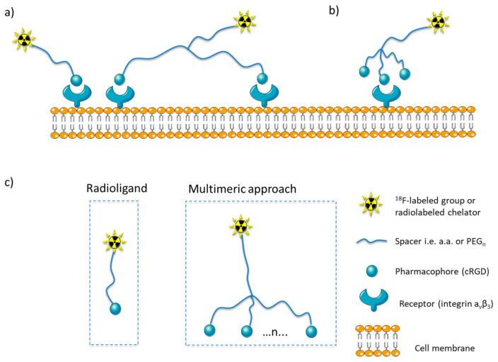
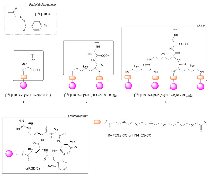
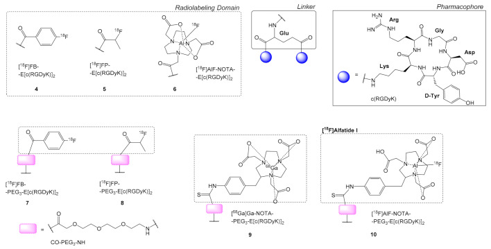
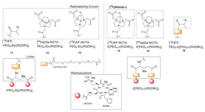
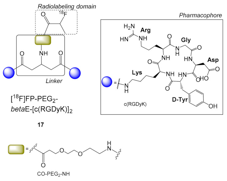
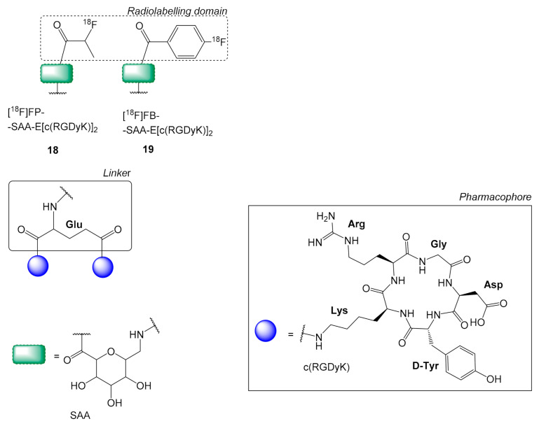
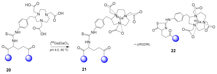
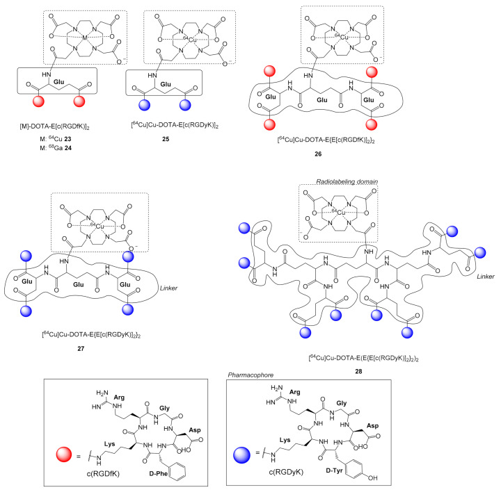
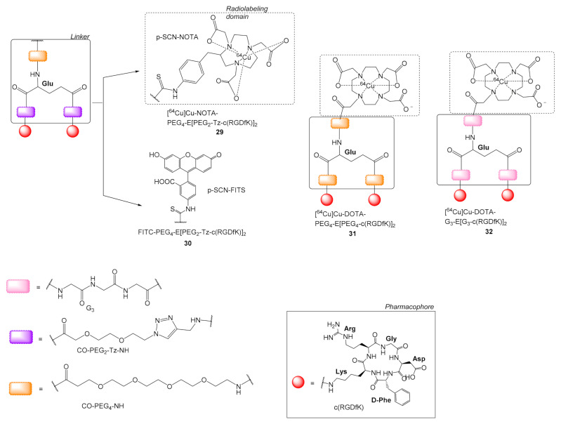
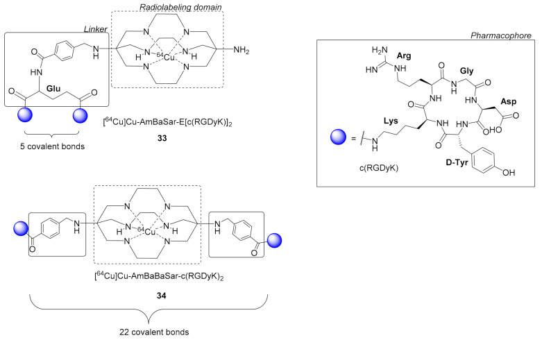
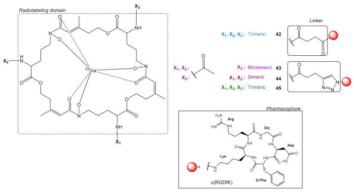
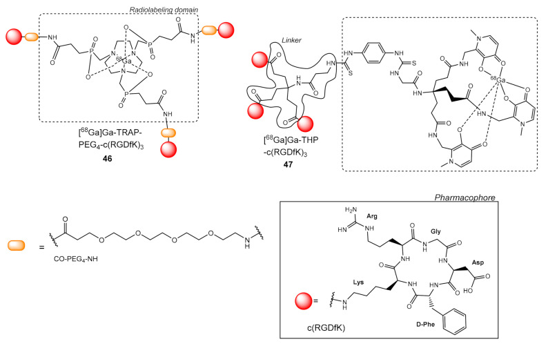
Multimeric ligands consisting of multiple pharmacophores connected to a single backbone have been widely investigated for diagnostic and therapeutic applications. In this review, we summarize recent developments regarding multimeric radioligands targeting integrin αvβ3 receptors on cancer cells for molecular imaging and diagnostic applications using positron emission tomography (PET). Integrin αvβ3 receptors are glycoproteins expressed on the cell surface, which have a significant role in tumor angiogenesis. They act as receptors for several extracellular matrix proteins exposing the tripeptide sequence arginine-glycine-aspartic (RGD). Cyclic RDG peptidic ligands c(RGD) have been developed for integrin αvβ3 tumor-targeting positron emission tomography (PET) diagnosis. Several c(RGD) pharmacophores, connected with the linker and conjugated to a chelator or precursor for radiolabeling with different PET radionuclides (18F, 64Cu, and 68Ga), have resulted in multimeric ligands superior to c(RGD) monomers. The binding avidity, pharmacodynamic, and PET imaging properties of these multimeric c(RGD) radioligands, in relation to their structural characteristics are analyzed and discussed. Furthermore, specific examples from preclinical studies and clinical investigations are included.

Keywords: PET imaging; cyclic RGD; integrin αvβ3; multimeric radioligands.


Conflict of interest statement

The authors declare no conflict of interest.

Figures

Figure 1
Figure 1
Binding models for multimers on the cell surface: (a) The binding of a radioligand to a cell surface receptors and the multimeric approach resulting in simultaneous binding of two pharmacophores connected via a long linker with two receptors, (b) improved binding efficiency of a ligand, due to the increased apparent local concentration of the pharmacophore (statistical effect) in the micro-environment of the receptor; (c) basic principles for the design of monomeric and multimeric radioligands (where n = number of pharmacophores).
Figure 2
Figure 2
Chemical structures of c(RGDfE) peptides with a PEG6 linker (H2N-PEG6-CH2COOH); monomer [18F]FBOA-Dpr-HEG-c(RGDfE) 1, dimer [18F]FBOA-Dpr-K(HEG-c(RGDfE))2 2 and tetramer [18F]FBOA-Dpr-K{K[HEG-(c(RGDfE)]2}2 3 are labeled with N-(4-[18F]fluorobenzylidene)oxime ([18F]FBOA) (Dpr = diaminopropionic acid).
Figure 3
Figure 3
Chemical structures of dimeric radioligands containing the scaffold E(c(RGDyK)]2 without: [18F]FB-E[c(RGDyK)]2 4, and [18F]FP-E[c(RGDyK)]2 5, [18F]AlF-NOTA-E[c(RGDyK)]2 6, and with a PEG3 group in between the alpha H2N- group of E and the radiolabeled domain: [18F]FB-PEG3-E[c(RGDfK)]2 7, [18F]FP-PEG3-E[c(RGDfK)]2 8, [68Ga]Ga-NOTA-PEG3-E[c(RGDyK)]2 9, [18F]AlF-NOTA-PEG3-E[c(RGDyK)]2 or [18F]Alfatide I 10 (H2N-PEG3-COOH = 11-amino-3,6,9-trioxaundecanoic acid).
Figure 4
Figure 4
Chemical structures of c(RGDfK)]2 analogues with PEG4 spacers; [18F]FP-PEG4-E[c(RGDfK)]2 11, [68Ga]Ga-NOTA-PEG4-E[c(RGDfK)]2 12, [18F]AlF-NOTA-PEG4-c(RGDfK)]2 13, [18F]AlF-NOTA-E[PEG4-c(RGDfK)]2 or [18F]Alfatide II 14, [68Ga]Ga-NOTA-E[PEG4-c(RGDfK)]2 15, [18F]FP-PEG4-E[PEG4-c(RGDfK)]2, 16 (HN-PEG4-COOH = 15-amino-4,7,10,13-tetraoxapentadecanoic acid).
Figure 5
Figure 5
Chemical structure of the symmetric dimer [18F]FP-PEG2-β-E[c(RGDyK)]2 17. (H2N-PEG2-COOH = 3-(2-(2-aminoethoxy)ethoxy)propanoic acid).
Figure 6
Figure 6
Chemical structures of [18F]FP-SAA-E[c(RGDyK)2 18, [18F]FB-SAA-E[c(RGDyK)2 19, where SAA = 7-amino-l-glyero-l-galacto-2,6-anhydro-7-deoxyheptanamide.
Figure 7
Figure 7
Instability of [68Ga]Ga-NOTA-E(c(RGDyK)]2 21 observed during its preparation from 20.
Figure 8
Figure 8
Chemical structures of 64Cu-labeled c(RGD) peptides with (Glu)n linkers, [M]-DOTA-E[c(RGDfK)]2, where M = 64Cu: 23, M = 68Ga: 24, [64Cu]Cu-DOTA-E[c(RGDyK)]2 25 and the c(RGD) tetramers [64Cu]Cu-DOTA-E{E[c(RGDfK)]2}2 26 and [64Cu]Cu-DOTA-E{E[c(RGDyK)]2}2 27 and the cRGD octamer [64Cu]Cu-DOTA-E(E{E[c(RGDfK)]2}2)2 28.
Figure 9
Figure 9
Chemical structures of 64Cu and FITC labeled c(RGD) peptides with the Tz-PEG2 spacers 29 and 30, respectively (Tz = triazole group, FITC = Fluorescein isothiocyanate isomer I) and with PEG4 31 and G3 32.
Figure 10
Figure 10
Dimeric c(RGDyK)2 radioligands [64Cu]Cu-AmBaSar-E[c(RGDyK)]2 32 and [64Cu]Cu-AmBaBaSar-c(RGDyK)2 33 bearing the hexaazamacrobicyclic sarcophagine (Sar) chelator, AmBa = 4-(Aminomethyl)benzoic acid.
Figure 11
Figure 11
Chemical structures of the dimeric radioligands [68Ga]Ga-NOTA-E[PEG4-c(RGDfK)]2 35, [68Ga]Ga-NOTA-E[G3-c(RGDfK)]2 36 and [68Ga]Ga-NOTA-E[G3-c(CNGRC)]2 37, [68Ga]Ga-NOTA-E[c(RGDyK)]2 38, [68Ga]Ga-NOTA-E{E[c(RGDyK)]2}2 39, [68Ga]Ga-NODAGA-E[c(RGDyK)]2 40 and [64Cu]Ga-NODAGA-E[c(RGDyK)]2 41.
Figure 12
Figure 12
Chemical structures of conjugates between the natural chelator fusarinine C (FSC) and c(RGDfK) pharmacophores, [68Ga]Ga-FSC-[E-c(RGDfK)]3 42, [68Ga]Ga-FSC-(CH2)-Tz-c(RGDfK) 43, [68Ga]Ga-FSC-[(CH2)-Tz-c(RGDfK)]2 44, [68Ga]Ga-FSC-[(CH2)-Tz-c(RGDfK)]3 45.
Figure 13
Figure 13
Chemical structures of 68Ga trimers of c(RGDfK) with TRAP 46 and THP 47 chelator groups.

References

    1. Handl H.L., Vagner J., Han H., Mash E., Hruby V.J., Gillies R.J. Hitting multiple targets with multimeric ligands. Expert Opin. Ther. Targets. 2004;8:565–586. doi: 10.1517/14728222.8.6.565. - DOI - PubMed
    1. Crothers D.M., Metzger H. The influence of polyvalency on the binding properties of antibodies. Immunochemistry. 1972;9:341–357. doi: 10.1016/0019-2791(72)90097-3. - DOI - PubMed
    1. Yim C.-B., Van Der Wildt B., Dijkgraaf I., Joosten L., Eek A., Versluis C., Rijkers D.T.S., Boerman O.C., Liskamp R.M.J. Spacer Effects on in vivo Properties of DOTA-Conjugated Dimeric [Tyr3]Octreotate Peptides Synthesized by a “CuI-Click” and “Sulfo-Click” Ligation Method. ChemBioChem. 2011;12:750–760. doi: 10.1002/cbic.201000639. - DOI - PubMed
    1. Baranski A.-C., Schäfer M., Bauder-Wüst U., Wacker A., Schmidt J., Liolios C., Mier W., Haberkorn U., Eisenhut M., Kopka K., et al. Improving the Imaging Contrast of 68Ga-PSMA-11 by Targeted Linker Design: Charged Spacer Moieties Enhance the Pharmacokinetic Properties. Bioconjugate Chem. 2017;28:2485–2492. doi: 10.1021/acs.bioconjchem.7b00458. - DOI - PubMed
    1. Liolios C.C., Fragogeorgi E.A., Zikos C., Loudos G., Xanthopoulos S., Bouziotis P., Paravatou-Petsotas M., Livaniou E., Varvarigou A.D., Sivolapenko G.B. Structural modifications of 99mTc-labelled bombesin-like peptides for optimizing pharmacokinetics in prostate tumor targeting. Int. J. Pharm. 2012;430:1–17. doi: 10.1016/j.ijpharm.2012.02.049. - DOI - PubMed

MeSH terms

Substances

LinkOut - more resources