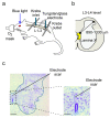
In Vivo Electrophysiology of Peptidergic Neurons in Deep Layers of the Lumbar Spinal Cord after Optogenetic Stimulation of Hypothalamic Paraventricular Oxytocin Neurons in Rats
- PMID: 33810239
- PMCID: PMC8036474
- DOI: 10.3390/ijms22073400
In Vivo Electrophysiology of Peptidergic Neurons in Deep Layers of the Lumbar Spinal Cord after Optogenetic Stimulation of Hypothalamic Paraventricular Oxytocin Neurons in Rats
Abstract
The spinal ejaculation generator (SEG) is located in the central gray (lamina X) of the rat lumbar spinal cord and plays a pivotal role in the ejaculatory reflex. We recently reported that SEG neurons express the oxytocin receptor and are activated by oxytocin projections from the paraventricular nucleus of hypothalamus (PVH). However, it is unknown whether the SEG responds to oxytocin in vivo. In this study, we analyzed the characteristics of the brain-spinal cord neural circuit that controls male sexual function using a newly developed in vivo electrophysiological technique. Optogenetic stimulation of the PVH of rats expressing channel rhodopsin under the oxytocin receptor promoter increased the spontaneous firing of most lamina X SEG neurons. This is the first demonstration of the in vivo electrical response from the deeper (lamina X) neurons in the spinal cord. Furthermore, we succeeded in the in vivo whole-cell recordings of lamina X neurons. In vivo whole-cell recordings may reveal the features of lamina X SEG neurons, including differences in neurotransmitters and response to stimulation. Taken together, these results suggest that in vivo electrophysiological stimulation can elucidate the neurophysiological response of a variety of spinal neurons during male sexual behavior.
Keywords: gastrin-releasing peptide neurons; in vivo extracellular recording; in vivo whole-cell patch-clamp recording; lamina X; optogenetics; oxytocin; spinal cord; spinal ejaculation generator.
Conflict of interest statement
The authors declare no conflict of interest.
Figures


Similar articles
-
Oxytocin Influences Male Sexual Activity via Non-synaptic Axonal Release in the Spinal Cord.Curr Biol. 2021 Jan 11;31(1):103-114.e5. doi: 10.1016/j.cub.2020.09.089. Epub 2020 Oct 29. Curr Biol. 2021. PMID: 33125871 Free PMC article.
-
Sexual Experience Induces the Expression of Gastrin-Releasing Peptide and Oxytocin Receptors in the Spinal Ejaculation Generator in Rats.Int J Mol Sci. 2021 Sep 26;22(19):10362. doi: 10.3390/ijms221910362. Int J Mol Sci. 2021. PMID: 34638701 Free PMC article.
-
Branched oxytocinergic innervations from the paraventricular hypothalamic nuclei to superficial layers in the spinal cord.Brain Res. 2007 Jul 30;1160:20-9. doi: 10.1016/j.brainres.2007.05.031. Epub 2007 May 26. Brain Res. 2007. PMID: 17599811
-
Neuropeptidergic control circuits in the spinal cord for male sexual behaviour: Oxytocin-gastrin-releasing peptide systems.J Neuroendocrinol. 2023 Sep;35(9):e13324. doi: 10.1111/jne.13324. Epub 2023 Jul 29. J Neuroendocrinol. 2023. PMID: 37515539 Review.
-
The role of oxytocin and the paraventricular nucleus in the sexual behaviour of male mammals.Physiol Behav. 2004 Nov 15;83(2):309-17. doi: 10.1016/j.physbeh.2004.08.019. Physiol Behav. 2004. PMID: 15488547 Review.
Cited by
-
Neurons for Ejaculation and Factors Affecting Ejaculation.Biology (Basel). 2022 Apr 29;11(5):686. doi: 10.3390/biology11050686. Biology (Basel). 2022. PMID: 35625414 Free PMC article. Review.
-
Developing a Novel Method for the Analysis of Spinal Cord-Penile Neurotransmission Mechanisms.Int J Mol Sci. 2023 Jan 11;24(2):1434. doi: 10.3390/ijms24021434. Int J Mol Sci. 2023. PMID: 36674942 Free PMC article.
-
Involvement of Histamine H3 Receptor Agonism in Premature Ejaculation Found by Studies in Rats.Int J Mol Sci. 2022 Feb 18;23(4):2291. doi: 10.3390/ijms23042291. Int J Mol Sci. 2022. PMID: 35216402 Free PMC article.
-
Near-Infrared Photobiomodulation of the Peripheral Nerve Inhibits the Neuronal Firing in a Rat Spinal Dorsal Horn Evoked by Mechanical Stimulation.Int J Mol Sci. 2023 Jan 25;24(3):2352. doi: 10.3390/ijms24032352. Int J Mol Sci. 2023. PMID: 36768673 Free PMC article.
-
Periostin activates distinct modules of inflammation and itching downstream of the type 2 inflammation pathway.Cell Rep. 2023 Jan 31;42(1):111933. doi: 10.1016/j.celrep.2022.111933. Epub 2023 Jan 6. Cell Rep. 2023. PMID: 36610396 Free PMC article.
References
MeSH terms
Substances
Grants and funding
LinkOut - more resources
Full Text Sources
Other Literature Sources

