Multimodal Imaging in Susac Syndrome: A Case Report and Literature Review
- PMID: 33810247
- PMCID: PMC8038062
- DOI: 10.3390/ijerph18073435
Multimodal Imaging in Susac Syndrome: A Case Report and Literature Review
Abstract
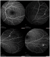
Susac syndrome (SS) is a rare microangiopathy that involves arterioles of the brain, retina, and cochlea. Diagnosis is extremely difficult because of the rarity of the disease and because the signs and symptoms often occur at different times. Multidisciplinary approaches and multimodal images are mandatory for diagnosis and prompt therapy. In this report, we describe a case of SS and the application of multimodal retinal imaging to evaluate the ophthalmologic changes and to confirm diagnosis. Early diagnosis and therapy based on the associations of steroids and immunosuppressants are necessary to limit the sequelae of the disease.
Keywords: fluorescein angiography; multimodal imaging; optical coherence tomography angiography; retinal branch artery occlusion; susac syndrome.
Conflict of interest statement
No potential conflicts of interest were reported by any author.
Figures


References
-
- Catarsi E., Pelliccia V., Pizzanelli C., Pesaresi I., Cosottini M., Migliorini P., Tavoni A. Cyclophosphamide and methotrexate in Susac’s Syndrome: A successful sequential therapy in a case with involvement of the cerebellum. Clin. Rheumatol. 2015;34:1149–1152. doi: 10.1007/s10067-014-2638-7. - DOI - PubMed
Publication types
MeSH terms
LinkOut - more resources
Full Text Sources
Other Literature Sources

