Review
. 2021 Mar 26;13(7):1027.
doi: 10.3390/polym13071027.

Recent Advancements in Polymer/Liposome Assembly for Drug Delivery: From Surface Modifications to Hybrid Vesicles

Affiliations
Review

Recent Advancements in Polymer/Liposome Assembly for Drug Delivery: From Surface Modifications to Hybrid Vesicles

Vincenzo De Leo et al. Polymers (Basel). .

Abstract

Liposomes are consolidated and attractive biomimetic nanocarriers widely used in the field of drug delivery. The structural versatility of liposomes has been exploited for the development of various carriers for the topical or systemic delivery of drugs and bioactive molecules, with the possibility of increasing their bioavailability and stability, and modulating and directing their release, while limiting the side effects at the same time. Nevertheless, first-generation vesicles suffer from some limitations including physical instability, short in vivo circulation lifetime, reduced payload, uncontrolled release properties, and low targeting abilities. Therefore, liposome preparation technology soon took advantage of the possibility of improving vesicle performance using both natural and synthetic polymers. Polymers can easily be synthesized in a controlled manner over a wide range of molecular weights and in a low dispersity range. Their properties are widely tunable and therefore allow the low chemical versatility typical of lipids to be overcome. Moreover, depending on their structure, polymers can be used to create a simple covering on the liposome surface or to intercalate in the phospholipid bilayer to give rise to real hybrid structures. This review illustrates the main strategies implemented in the field of polymer/liposome assembly for drug delivery, with a look at the most recent publications without neglecting basic concepts for a simple and complete understanding by the reader.

Keywords: drug release profile; encapsulation efficiency; hybrid vesicles; liposome surface modification; liposomes; mucopenetrating/mucoadhesive properties; physicochemical stability; polymers; stimuli-responsive properties; versatile targeting platform.


Conflict of interest statement

The authors declare no conflict of interest.

Figures

Scheme 1
Scheme 1
Chemical structures of most commonly used (phospho)lipids for liposome preparation. From top to bottom: The zwitterionic 1-palmitoyl-2-oleoyl-phosphatidylcholine (POPC), principal constituent of natural membranes; the zwitterionic and fusogenic 1-palmitoyl-2-oleoyl-phosphatidylethanolamine (POPE); the anionic 1-palmitoyl-2-oleoyl-phosphatidylglycerol (POPG); the cationic lipid 1,2-dioleoyl-3-trimethylammonium-propane (DOTAP). Bottom: Schematic drawing of a generic phospholipid with the two alkyl chains depicted as black wires and the polar head represented by a red circle.
Figure 1
Figure 1
Polymer/liposome assembly. (A) Hydrophilic polymers are physically adsorbed onto liposome surface; (B) polymers are polymerized or undergo a phase change, trapping the vesicles inside; (C, left) polymers are covalently bonded to the polar head of phospholipids; (C, right) polymerizable monomers are reticulated to form a network surrounding the vesicles; (D, left) amphiphilic polymers are homogeneously distributed in the bilayer or (D, right) segregated in separate domains.
Figure 2
Figure 2
Scheme representing the mechanism for increased circulation time of PEG-decorated liposomes. (A) Conventional liposomes, bearing hydrophilic (orange balls) and hydrophobic (yellow stars) loads, aggregated and attacked by plasma proteins (Y-shaped antibodies in yellow and green); (B) stealth liposomes with PEG functionalization (cyan “hairs”) whose surface cannot be reached by plasma proteins.
Figure 3
Figure 3
Hybrid stealth liposomes stabilized by cholesteryl-functionalized block copolymers. The cholesteryl moieties (red sticks) insert into the bilayer while the tails (in blue) protrude toward the aqueous phase, protecting the liposomes from coalescence and plasma proteins. Reproduced with permission from [52]. Copyright © 2018 American Chemical Society.
Figure 4
Figure 4
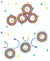
(A) Conventional liposome-bearing hydrophilic (orange balls) and hydrophobic (yellow stars) loads. The hydrophilic cargo tends to cross the bilayer leaking to the external aqueous phase. (B) Polymer-coated liposome with reduced bilayer permeability and higher cargo retention, enabling sustained and controlled drug delivery.
Figure 5
Figure 5
Scheme of a generic tissue formed by several cells (in orange with blue nuclei) covered by mucus (in yellow). (A) Conventional liposome unable to cross the mucosal barrier; (B) liposome covered by polymer, increasing the mucoadhesive properties of the vesicle; (C) liposome decorated with polymer conferring mucopenetrating properties to the vesicle.
Figure 6
Figure 6
Maleimide-functionalized PEGylated liposomes showing mucoadhesive properties thanks to the covalent linkages with thiol-groups present in mucins. Reproduced with permission from [97]. Copyright © 2018 Elsevier.
Figure 7
Figure 7
Experimental design to assess the mucopenetrative properties of plain-, PF127-, and PEG-liposomes, using the mucus of chronic obstructive pulmonary disease (COPD) patients. Reproduced with permission from [22]. Copyright © 2018 Elsevier.
Figure 8
Figure 8
Stimuli-responsive polymeric coatings of liposomes. In (A), the stimulus dissolves the protecting coating, while in (B), the stimulus destabilizes the bilayer and/or induces the formation of transmembrane channels.
Figure 9
Figure 9
Design of temperature and pH dual-stimuli-responsive liposomes using hyperbranched poly(glycidol)s with temperature-sensitive OEG groups and pH-sensitive succinyl groups. Reproduced with permission from [112]. Copyright © 2014 Elsevier.
Figure 10
Figure 10
Schematic illustration of liposome containing doxorubicin and rapamycin and complexed with glycol chitosan. The nanovectors are stable at pH 7.4 and aggregate when the pH is lowered to 5.0. Reproduced under Creative Common license from ref [121].
Figure 11
Figure 11
Schematic representation of targeted liposomes, with targeting ligands covalently linked to the distal end of PEG-lipids. For illustrative purposes, antibodies are shown on the left, aptamer and sugar in the middle, and a generic small molecule and peptide are sketched on the right.

References

    1. Walde P. Preparation of Vesicles (Liposomes) In: Nalwa H.S., editor. Encyclopedia of Nanoscience and Nanotechnology. Vol. 8. American Scientific Publishers; Valencia, CA, USA: 2003. pp. 1–37.
    1. Gregoriadis G. Liposome Preparation and Related Techniques. CRC Press; Boca Raton, FL, USA: 2006.
    1. New R. Liposomes: A Practical Approach. IRL PRESS; New York, NY, USA: Oxford University Press; Oxford, UK: 1990.
    1. Stano P., D’Aguanno E., Bolz J., Fahr A., Luisi P.L. A Remarkable Self-Organization Process as the Origin of Primitive Functional Cells. Angew. Chem. Int. Ed. 2013;52:13397–13400. doi: 10.1002/anie.201306613. - DOI - PubMed
    1. Sercombe L., Veerati T., Moheimani F., Wu S.Y., Sood A.K., Hua S. Advances and Challenges of Liposome Assisted Drug Delivery. Front. Pharmacol. 2015;6:286. doi: 10.3389/fphar.2015.00286. - DOI - PMC - PubMed

LinkOut - more resources