. 2021 Mar 26;10(4):264.
doi: 10.3390/biology10040264.

Kaempferia parviflora Rhizome Extract Inhibits Glutamate-Induced Toxicity in HT-22 Mouse Hippocampal Neuronal Cells and Extends Longevity in Caenorhabditis elegans

Affiliations

Kaempferia parviflora Rhizome Extract Inhibits Glutamate-Induced Toxicity in HT-22 Mouse Hippocampal Neuronal Cells and Extends Longevity in Caenorhabditis elegans

Aunchalee Tonsomboon et al. Biology (Basel). .

Abstract

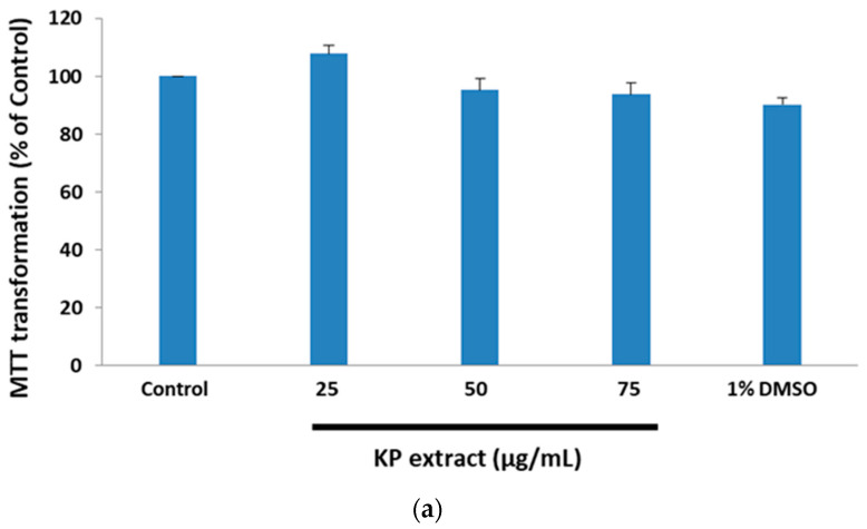
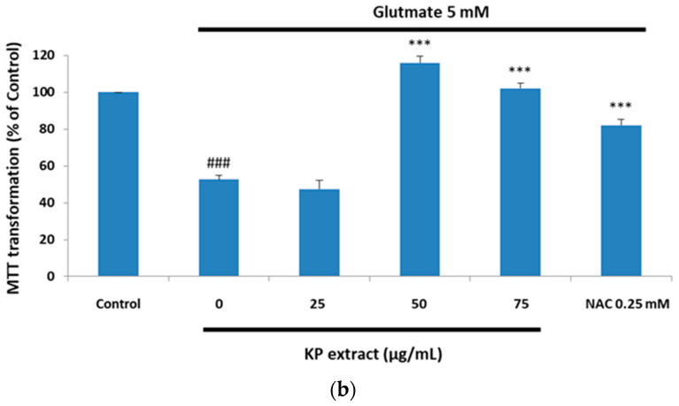
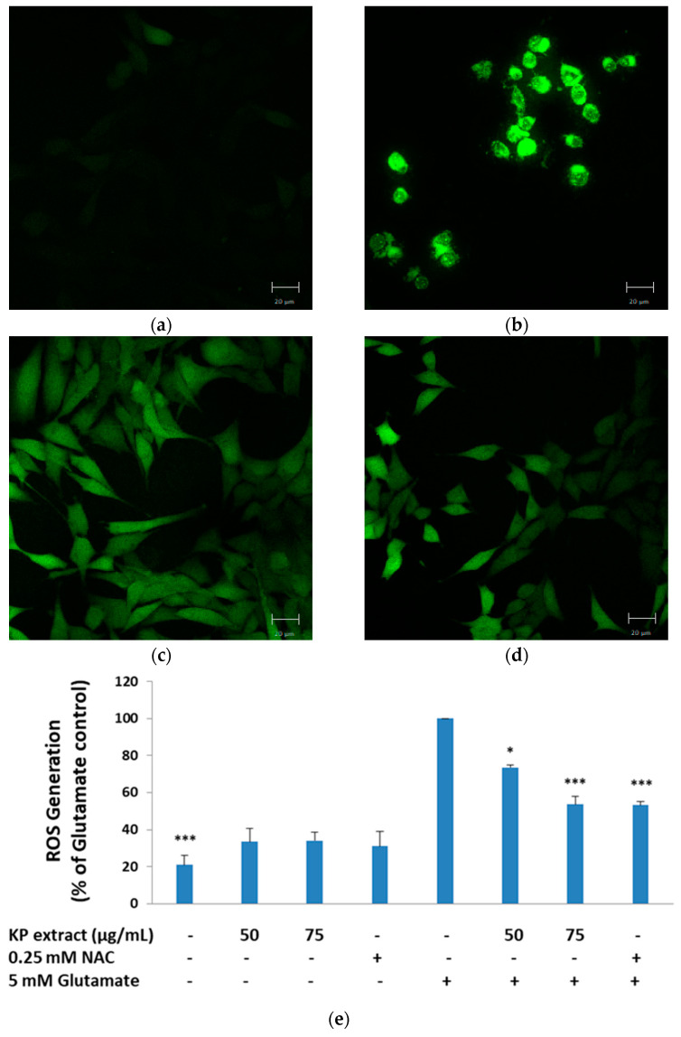
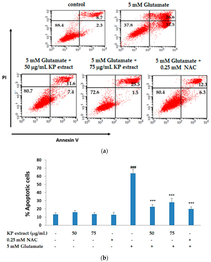
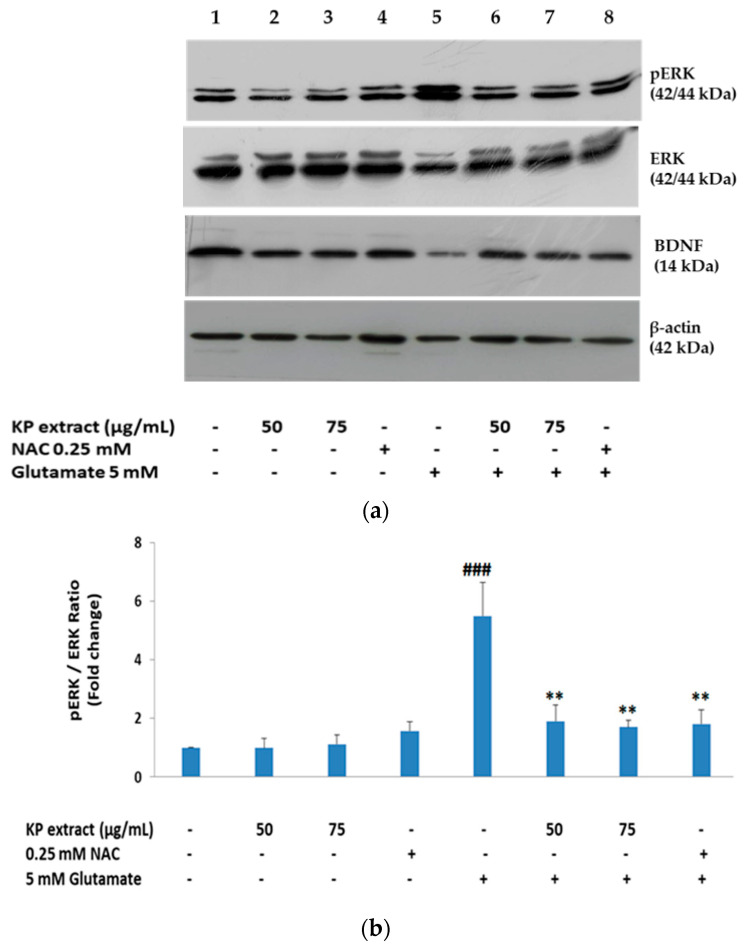
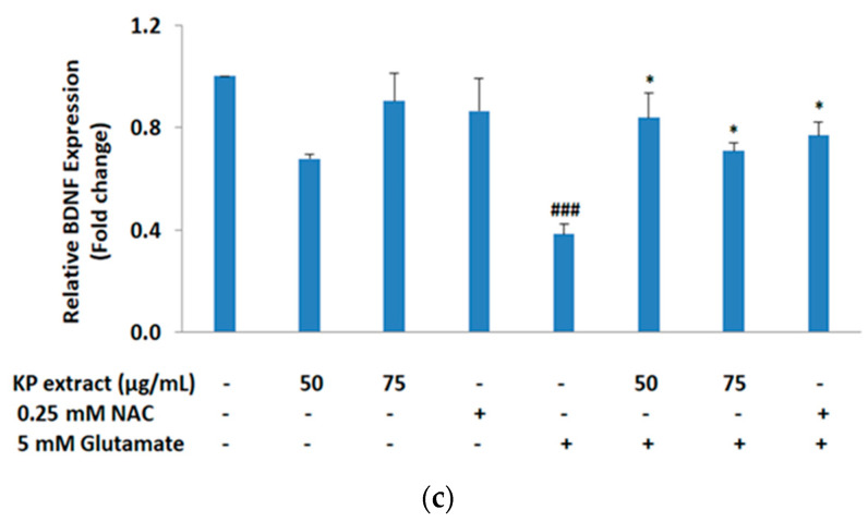
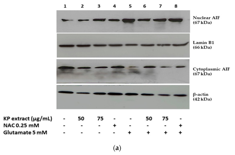
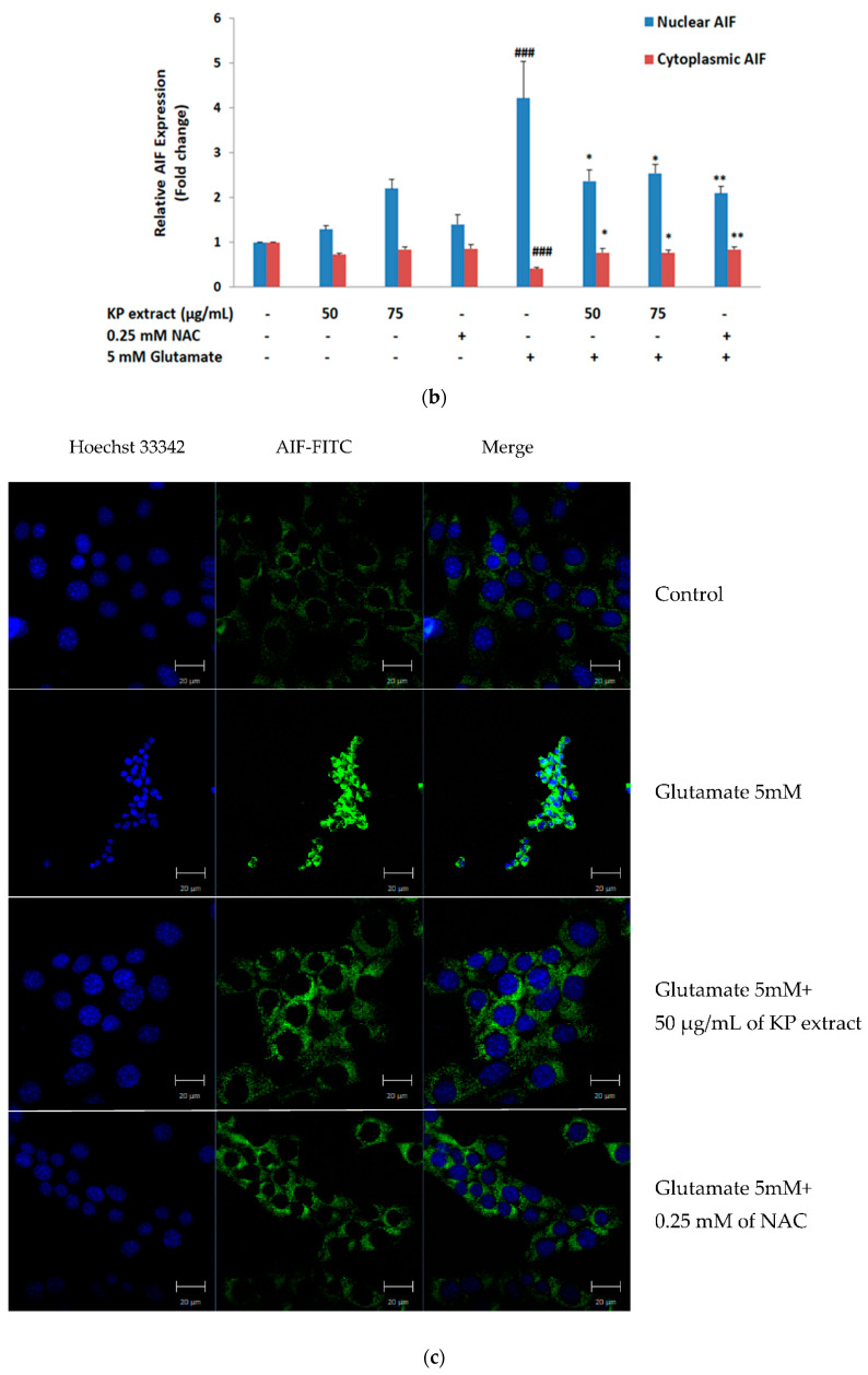
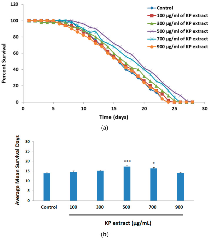
Kaempferia parviflora Wall. ex Baker (KP) or "Kra-chai-dam" has been shown to exhibit several pharmacological effects including anti-inflammation, antimicrobial, and sexual-enhancing activity. The objectives of this study included an investigation of the effect of KP rhizome extract against glutamate-induced toxicity in mouse hippocampal HT-22 neuronal cells, determination of the underlying mechanism of neuroprotection, and an evaluation of the effect of KP extract on the longevity of Caenorhabditis elegans. HT-22 cells were co-treated with glutamate (5 mM) and KP extract (25, 50, and 75 μg/mL) for 14 h. Cell viability, intracellular reactive oxygen species (ROS) assay, fluorescence-activated cell sorting (FACS) analysis, and Western blotting were performed. The longevity effect of KP extract on C. elegans was studied by lifespan measurement. In HT-22 cells, co-treatment of glutamate with KP extract significantly inhibited glutamate-mediated cytotoxicity and decreased intracellular ROS production. Additionally, the glutamate-induced apoptosis and apoptotic-inducing factor (AIF) translocation were blocked by KP extract co-treatment. Western blot analysis also demonstrated that KP extract significantly diminished extracellular signal-regulated kinase (ERK) phosphorylation induced by glutamate, and brain-derived neurotrophic factor (BDNF) was recovered to the control. Moreover, this KP extract treatment prolonged the lifespan of C. elegans. Altogether, this study suggested that KP extract possesses both neuroprotective and longevity-inducing properties, thus serving as a promising candidate for development of innovative health products.

Keywords: Caenorhabditis elegans; HT-22 mouse hippocampal neuronal cells; Kaempferia parviflora rhizome extract; glutamate toxicity.


Conflict of interest statement

The authors declare no conflict of interest.

Figures

Figure 1
Figure 1
Neuroprotective effect of Kaempferia parviflora Wall. ex Baker (KP) extract on glutamate-induced cytotoxicity in HT-22 cells. (a) Cells were treated with various concentrations of KP extract (25, 50, and 75 µg/mL) for 14 h; (b) cells were exposed to 5 mM of glutamate alone or glutamate in combination with different concentrations of KP extract for 14 h. Cell viability was determined by 3-(4,5-dimethylthiazol-2-yl)- 2,5-diphenyl tetrazolium bromide (MTT) assay. Each bar represents the mean ± SEM from 3 independent experiments per group. ### p < 0.001 vs. control and *** p < 0.001 vs. the glutamate alone.
Figure 1
Figure 1
Neuroprotective effect of Kaempferia parviflora Wall. ex Baker (KP) extract on glutamate-induced cytotoxicity in HT-22 cells. (a) Cells were treated with various concentrations of KP extract (25, 50, and 75 µg/mL) for 14 h; (b) cells were exposed to 5 mM of glutamate alone or glutamate in combination with different concentrations of KP extract for 14 h. Cell viability was determined by 3-(4,5-dimethylthiazol-2-yl)- 2,5-diphenyl tetrazolium bromide (MTT) assay. Each bar represents the mean ± SEM from 3 independent experiments per group. ### p < 0.001 vs. control and *** p < 0.001 vs. the glutamate alone.
Figure 2
Figure 2
Inhibitory effect of KP extract against glutamate-induced oxidative stress in HT-22 cells. HT-22 cells were treated with 5 mM glutamate alone or co-treatment of glutamate with various concentrations of KP extract for 14 h. The production of intracellular reactive oxygen species (ROS) was stained by 2′, 7′-dichlorodihydro fluorescein diacetate (H2DCFDA) and observed under a fluorescence microscope. (a) Untreated cells (control); (b) 5 mM of glutamate; (c) 75 µg/mL of KP extract + 5 mM of glutamate; (d) 0.25 mM of N-acetyl cysteine (NAC) + 5 mM of glutamate; (e) the quantity of production of intracellular ROS was measured using H2DCFDA by the fluorescence microplate reader. All data are shown as the mean ± SEM of at least three independent experiments. * p < 0.05 and *** p < 0.001 vs. the glutamate treatment alone. The symbol “-” and “+” are defined as absent and present, respectively.
Figure 3
Figure 3
The anti-apoptotic activity of KP extract on HT-22 cells. (a) Representative flow cytometry plots using Annexin V-FITC/PI staining in each group including an untreated group (control), 50 and 75 µg/mL of KP extract treatment in the presence or absence of 5 mM of glutamate for 14 h. NAC (0.25 mM) was used as positive control; (b) bar graph indicates the percentage of apoptotic cells (PI-/annexin V + stained cell population). Data are presented as the mean ± SEM (n = 3). ### p < 0.001 vs. control and *** p < 0.001 vs. 5 mM of glutamate. The symbol “-” and “+” are defined as absent and present, respectively.
Figure 4
Figure 4
Effect of KP extract on the glutamate-induced phosphorylation of extracellular signal- regulated kinase (ERK) and brain-derived neurotrophic factor (BDNF) expression. (a) HT-22 cells were exposed with 50 and 75 μg/mL KP extract in the presence or absence of glutamate (5 mM) for 14 h, NAC (0.25 mM) was used as positive control. Following incubation, the cells were harvested and BDNF, ERK, and protein kinase-like endoplasmic reticulum kinase (p-ERK) levels were investigated by Western blot analysis. The immunoreactive bands were detected using specific antibodies for BDNF, ERK, p-ERK, and β-actin; (b,c) bars represent the fold-increase in phosphorylation of mitogen-activated protein kinases (MAPKs) and BDNF expression, respectively. All data were normalized to internal control levels and are expressed as the mean ± SEM (n = 3), ### p < 0.001 vs. control, * p < 0.05 and ** p < 0.01 vs. glutamate alone. The symbol “-” and “+” are defined as absent and present, respectively.
Figure 4
Figure 4
Effect of KP extract on the glutamate-induced phosphorylation of extracellular signal- regulated kinase (ERK) and brain-derived neurotrophic factor (BDNF) expression. (a) HT-22 cells were exposed with 50 and 75 μg/mL KP extract in the presence or absence of glutamate (5 mM) for 14 h, NAC (0.25 mM) was used as positive control. Following incubation, the cells were harvested and BDNF, ERK, and protein kinase-like endoplasmic reticulum kinase (p-ERK) levels were investigated by Western blot analysis. The immunoreactive bands were detected using specific antibodies for BDNF, ERK, p-ERK, and β-actin; (b,c) bars represent the fold-increase in phosphorylation of mitogen-activated protein kinases (MAPKs) and BDNF expression, respectively. All data were normalized to internal control levels and are expressed as the mean ± SEM (n = 3), ### p < 0.001 vs. control, * p < 0.05 and ** p < 0.01 vs. glutamate alone. The symbol “-” and “+” are defined as absent and present, respectively.
Figure 5
Figure 5
The effect of KP extract against glutamate-mediated cell death involves apoptotic-inducing factor (AIF) translocation to the nucleus. HT-22 cells were exposed with 50 and 75 μg/mL KP extract in the presence or absence of glutamate (5 mM) for 14 h, NAC (0.25 mM) was used as positive control. (a) Following incubation, nuclear and cytoplasmic fractionation was performed and all targeted proteins (then, nuclear AIF, cytoplasmic AIF, Lamin B1, and β-actin were analyzed using Western blot analysis; (b) bars represent the fold-change of nuclear and cytoplasmic AIF expression. Lamin B1 and β-actin were used as an internal control to normalize protein expression for nuclear extracts and whole cell/cytoplasmic extracts, respectively. All data are expressed as the mean ± SEM (n = 3), ### p < 0.001 vs. control, * p < 0.05, and ** p < 0.01 vs. glutamate alone. The symbol “-” and “+” are defined as absent and present, respectively; (c) confocal laser scanning microscope images of AIF immunoreactivity (green) and nuclear Hoechst 33342 staining (dark blue). Co-treatment with KP extract resulting in prevention of nuclear AIF translocation after 14 h of 5 mM of glutamate exposure.
Figure 5
Figure 5
The effect of KP extract against glutamate-mediated cell death involves apoptotic-inducing factor (AIF) translocation to the nucleus. HT-22 cells were exposed with 50 and 75 μg/mL KP extract in the presence or absence of glutamate (5 mM) for 14 h, NAC (0.25 mM) was used as positive control. (a) Following incubation, nuclear and cytoplasmic fractionation was performed and all targeted proteins (then, nuclear AIF, cytoplasmic AIF, Lamin B1, and β-actin were analyzed using Western blot analysis; (b) bars represent the fold-change of nuclear and cytoplasmic AIF expression. Lamin B1 and β-actin were used as an internal control to normalize protein expression for nuclear extracts and whole cell/cytoplasmic extracts, respectively. All data are expressed as the mean ± SEM (n = 3), ### p < 0.001 vs. control, * p < 0.05, and ** p < 0.01 vs. glutamate alone. The symbol “-” and “+” are defined as absent and present, respectively; (c) confocal laser scanning microscope images of AIF immunoreactivity (green) and nuclear Hoechst 33342 staining (dark blue). Co-treatment with KP extract resulting in prevention of nuclear AIF translocation after 14 h of 5 mM of glutamate exposure.
Figure 6
Figure 6
Effect of KP extracts on the lifespan of N2 wild-type C. elegans. The nematodes were treated with various concentration of KP extracts (0, 100, 300, 500, 700, and 900 µg/mL) at 15 °C. The survival was counted every day till death. (a) Survival curves of KP extracted treated C. elegans as compared with the untreated group (control); (b) Bars represent the mean lifespan when treated with KP extract. The experiment was performed 5 independent trails and shown as the mean ± SEM, * p < 0.05 and *** p < 0.001 vs. control.

Similar articles

Cited by

References

    1. Luu J., Palczewski K. Human aging and disease: Lessons from age-related macular degeneration. Proc. Natl. Acad. Sci. USA. 2018;115:2866–2872. doi: 10.1073/pnas.1721033115. - DOI - PMC - PubMed
    1. Alzheimer’s Association 2016 Alzheimer’s disease facts and figures. Alzheimers Dement. 2016;12:459–509. doi: 10.1016/j.jalz.2016.03.001. - DOI - PubMed
    1. World Health Organization Thailand: Alzheimers/Dementia 2017. [(accessed on 10 July 2020)];2017 Available online: https://www.worldlifeexpectancy.com/thailand-alzheimers-dementia.
    1. Kritis A.A., Stamoula E.G., Paniskaki K.A., Vavilis T.D. Researching glutamate—Induced cytotoxicity in different cell lines: A comparative/collective analysis/study. Front. Cell. Neurosci. 2015;9:91. doi: 10.3389/fncel.2015.00091. - DOI - PMC - PubMed
    1. Joseph T.C., Pamela P. Oxidative Stress, Glutamate, and Neurodegenerative Disorders. Science. 1993;262:689–695. - PubMed

LinkOut - more resources