

Eltrombopag Improves Erythroid Differentiation in a Human Induced Pluripotent Stem Cell Model of Diamond Blackfan Anemia
- PMID: 33810313
- PMCID: PMC8065708
- DOI: 10.3390/cells10040734
Eltrombopag Improves Erythroid Differentiation in a Human Induced Pluripotent Stem Cell Model of Diamond Blackfan Anemia
Abstract
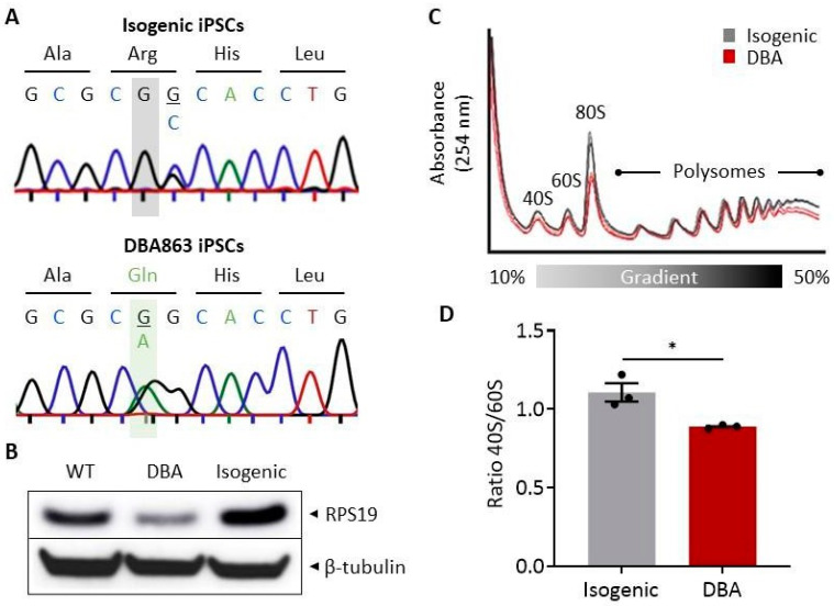
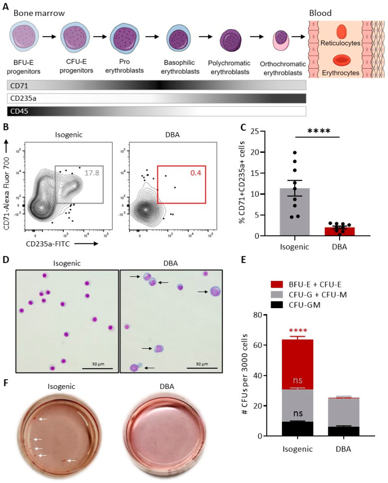
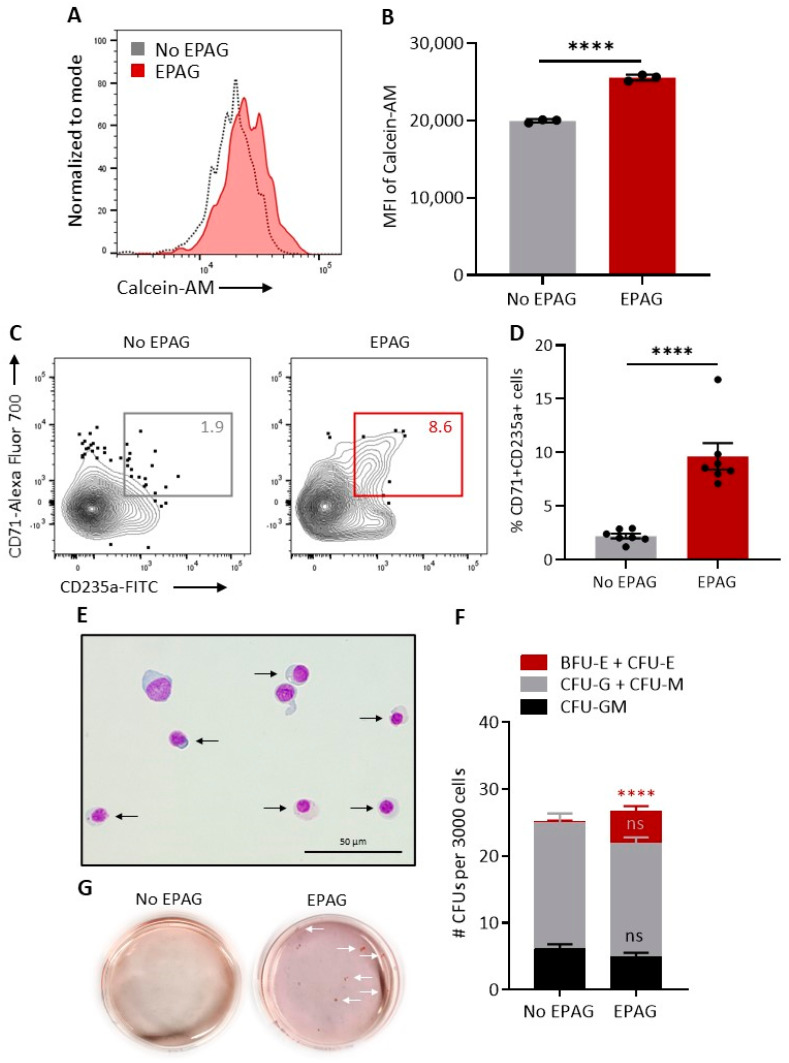
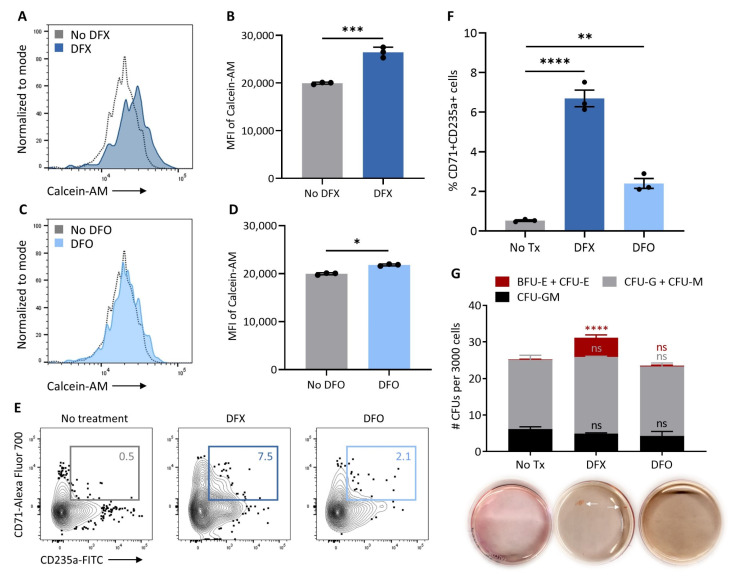
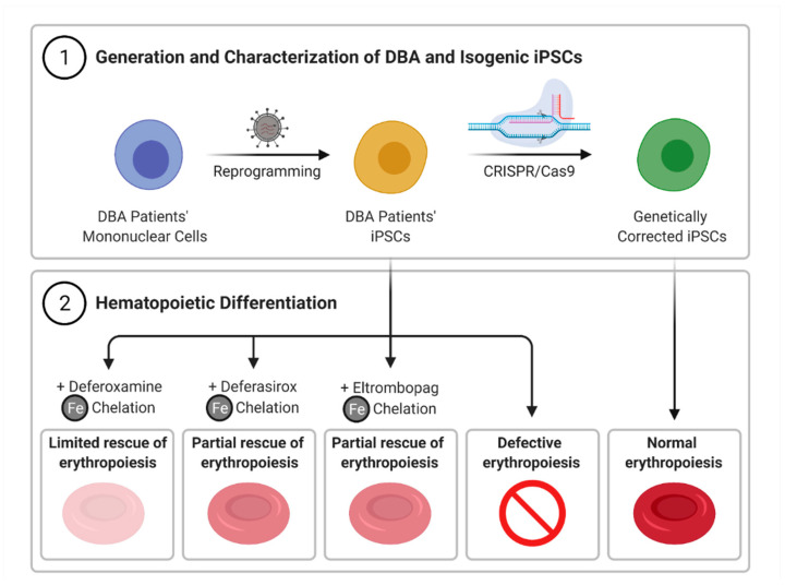
Diamond Blackfan Anemia (DBA) is a congenital macrocytic anemia associated with ribosomal protein haploinsufficiency. Ribosomal dysfunction delays globin synthesis, resulting in excess toxic free heme in erythroid progenitors, early differentiation arrest, and pure red cell aplasia. In this study, DBA induced pluripotent stem cell (iPSC) lines were generated from blood mononuclear cells of DBA patients with inactivating mutations in RPS19 and subjected to hematopoietic differentiation to model disease phenotypes. In vitro differentiated hematopoietic cells were used to investigate whether eltrombopag, an FDA-approved mimetic of thrombopoietin with robust intracellular iron chelating properties, could rescue erythropoiesis in DBA by restricting the labile iron pool (LIP) derived from excessive free heme. DBA iPSCs exhibited RPS19 haploinsufficiency, reduction in the 40S/60S ribosomal subunit ratio and early erythroid differentiation arrest in the absence of eltrombopag, compared to control isogenic iPSCs established by CRISPR/Cas9-mediated correction of the RPS19 point mutation. Notably, differentiation of DBA iPSCs in the presence of eltrombopag markedly improved erythroid maturation. Consistent with a molecular mechanism based on intracellular iron chelation, we observed that deferasirox, a clinically licensed iron chelator able to permeate into cells, also enhanced erythropoiesis in our DBA iPSC model. In contrast, erythroid maturation did not improve substantially in DBA iPSC differentiation cultures supplemented with deferoxamine, a clinically available iron chelator that poorly accesses LIP within cellular compartments. These findings identify eltrombopag as a promising new therapeutic to improve anemia in DBA.
Trial registration: ClinicalTrials.gov NCT00027274.
Keywords: diamond blackfan anemia; disease modeling; drug testing; eltrombopag; genome editing; induced pluripotent stem cells.
Conflict of interest statement
STEMdiff™ Hematopoietic Kit used in this study is registered under patent WO2015050963 A1. AL receives royalty income. Novartis, the manufacturer of eltrombopag, provided research grade drug. The other authors declare no competing interests.
Figures
Similar articles
-
Treatment of refractory/relapsed Diamond-Blackfan anaemia with eltrombopag.Br J Haematol. 2024 May;204(5):2077-2085. doi: 10.1111/bjh.19357. Epub 2024 Mar 10. Br J Haematol. 2024. PMID: 38462764 Free PMC article. Clinical Trial.
-
Drug discovery for Diamond-Blackfan anemia using reprogrammed hematopoietic progenitors.Sci Transl Med. 2017 Feb 8;9(376):eaah5645. doi: 10.1126/scitranslmed.aah5645. Sci Transl Med. 2017. PMID: 28179501 Free PMC article.
-
Regulation of globin-heme balance in Diamond-Blackfan anemia by HSP70/GATA1.Blood. 2019 Mar 21;133(12):1358-1370. doi: 10.1182/blood-2018-09-875674. Epub 2019 Jan 30. Blood. 2019. PMID: 30700418 Free PMC article.
-
Diamond-Blackfan anemia: erythropoiesis lost in translation.Blood. 2007 Apr 15;109(8):3152-4. doi: 10.1182/blood-2006-09-001222. Epub 2006 Dec 12. Blood. 2007. PMID: 17164339 Review.
-
Recent insights into the pathogenesis of Diamond-Blackfan anaemia.Br J Haematol. 2006 Oct;135(2):149-57. doi: 10.1111/j.1365-2141.2006.06268.x. Epub 2006 Aug 31. Br J Haematol. 2006. PMID: 16942586 Review.
Cited by
-
A De Novo Frameshift Mutation in RPL5 with Classical Phenotype Abnormalities and Worsening Anemia Diagnosed in a Young Adult-A Case Report and Review of the Literature.Medicina (Kaunas). 2023 Nov 5;59(11):1953. doi: 10.3390/medicina59111953. Medicina (Kaunas). 2023. PMID: 38004002 Free PMC article. Review.
-
Editorial for Special Issue: iPS Cells (iPSCs) for Modelling and Treatment of Human Diseases.Cells. 2022 Jul 22;11(15):2270. doi: 10.3390/cells11152270. Cells. 2022. PMID: 35892567 Free PMC article.
-
Modulation of WNT, Activin/Nodal, and MAPK Signaling Pathways Increases Arterial Hemogenic Endothelium and Hematopoietic Stem/Progenitor Cell Formation During Human iPSC Differentiation.Stem Cells. 2023 Jul 14;41(7):685-697. doi: 10.1093/stmcls/sxad040. Stem Cells. 2023. PMID: 37220178 Free PMC article.
-
Studies of a mosaic patient with DBA and chimeric mice reveal erythroid cell-extrinsic contributions to erythropoiesis.Blood. 2022 Jun 9;139(23):3439-3449. doi: 10.1182/blood.2021013507. Blood. 2022. PMID: 35349664 Free PMC article.
-
Treatment of refractory/relapsed Diamond-Blackfan anaemia with eltrombopag.Br J Haematol. 2024 May;204(5):2077-2085. doi: 10.1111/bjh.19357. Epub 2024 Mar 10. Br J Haematol. 2024. PMID: 38462764 Free PMC article. Clinical Trial.
References
-
- Payne E.M., Virgilio M., Narla A., Sun H., Levine M., Paw B.H., Berliner N., Look A.T., Ebert B.L., Khanna-Gupta A. L-leucine improves the anemia and developmental defects associated with Diamond-Blackfan anemia and del(5q) MDS by activating the mTOR pathway. Blood. 2012;120:2214–2224. doi: 10.1182/blood-2011-10-382986. - DOI - PMC - PubMed
Publication types
MeSH terms
Substances
Associated data
Grants and funding
LinkOut - more resources
Full Text Sources
Other Literature Sources
Medical
Miscellaneous
