. 2021 Mar 26;11(4):940.
doi: 10.3390/ani11040940.

Gene Expression Profile in Peripheral Blood Nuclear Cells of Small Ruminant Lentivirus-Seropositive and Seronegative Dairy Goats in Their First Lactation

Affiliations

Gene Expression Profile in Peripheral Blood Nuclear Cells of Small Ruminant Lentivirus-Seropositive and Seronegative Dairy Goats in Their First Lactation

Joanna Pławińska-Czarnak et al. Animals (Basel). .

Abstract

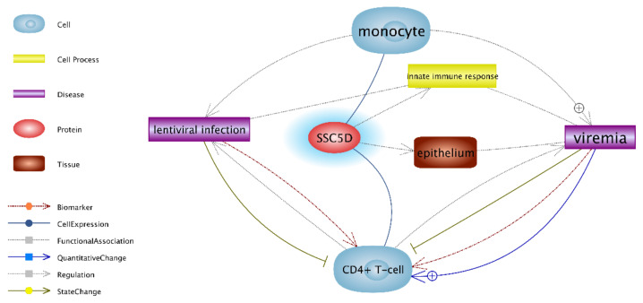
The immune response to a viral antigen causes inflammatory cell infiltration to the tissue, which creates a suitable environment for the replication of the virus in macrophages, and the recruitment of more monocytes to the site of infection, or latently infected monocytes. The aim of the study was to analyze the transcriptomic profile of peripheral blood nuclear cells isolated from SRLV-seropositive and SRLV-negative goats at the peak of their first lactation. SRLV-seropositive goats were probably infected via colostrum. Custom transcriptomic microarrays for goats were designed and developed, namely the Capra hircus gene expression array, which features ~50,000 unique transcripts per microarray. Only four genes were differentially expressed, with up-regulated expression of the GIMAP2, SSC5D and SETX genes, and down-regulated expression of the GPR37 gene in SRLV-seropositive vs. SRLV-seronegative goats. However, in an RT-qPCR analysis, the result for the SETX gene was not confirmed. The differences in the expressions of the studied genes indicate an active inflammatory process in the SRLV-seropositive goats at the early stage of infection.

Keywords: GIMAP2; GPR37; SETX; SSC5D; gene expression; goat; microarray; peripheral blood nuclear cells; small ruminant lentivirus.


Conflict of interest statement

The authors declare no conflict of interest. The funders had no role in the design of the study; in the collection, analyses, or interpretation of data; in the writing of the manuscript, or in the decision to publish the results.

Figures

Figure 1
Figure 1
Direct links between the SSC5D protein and monocytes and CD4+ T cells during lentivirus infection. Upregulated expression of the SSC5D gene in PBNCs of SRLV-SP vs. SRLV-SN goats, as analyzed by the Pathway Studio program [30]. The blue line highlights the upregulation of the gene in SRLV-SP goats vs. SRLV-SN goats. SSC5D—scavenger receptor cysteine-rich family member with five domains (alias S5D-SRCRB); PBNCs—peripheral blood nuclear cells; RT-qPCR—quantitative reverse transcription PCR; SRLV-SP—small ruminant lentivirus-seropositive; SRLV-SN—small ruminant lentivirus-seronegative.
Figure 2
Figure 2
Interactions between proteins, cells, and lentiviral infection for the GIMAP2 gene in PBNCs, as analyzed by the Pathway Studio program [30]. Blue highlights upregulation in SRLV-SP goats vs. SRLV-SN goats; GIMAP2—GTPase, IMAP family member 2; GIMAP7—GTPase, IMAP family member 7 (GIMAP7 -> GIMAP2 can stimulate both its own GTPase activity and that of GIMAP2 by dimerization and the provision of a catalytic arginine finger); DNM1L—dynamin-1-like protein (gene encodes a member of the dynamin superfamily of GTPases); SLC7A7—solute carrier family 7 member 7 (regulates 11 gene expression signature genes, including GIMAP2, IL10RA, and IL18as); IL7R—interleukin 7 receptors (->Regulation IL7R->GIMAP2; the signal given by IL-7/IL-7R affects the regulation of apoptosis, cell cycle progression, and differentiation, cell type CD4+ T cell); GDP—guanosine diphosphate; GTP—guanosine triphosphate; IL7—interleukin 7; CD4+ T-cell—T-cell surface glycoprotein CD4 precursor (this gene encodes a membrane glycoprotein of T lymphocytes that interacts with major histocompatibility complex class II antigens and is also a receptor for the human immunodeficiency virus).
Figure 3
Figure 3
Involvement of GPR37 in processes related to the cellular response to infection with small ruminant lentiviruses/visna-medi virus in goats. Pink highlights downregulation; GPR37—G protein-coupled receptor 37; FOS-Fos proto-oncogene, (AP-1 transcription factor subunit) is a protein coding gene. These genes encode leucine zipper proteins that can dimerize with proteins of the JUN family, thereby forming the AP-1 transcription factor complex. The interaction between Fos and Jun targets the Tat visna virus and promotes its replication in monocytes and macrophages after their differentiation. Fos-Jun activation can influence the activation of many cellular genes; this could intensely affect cell and viral growth and thus play a role in the pathogenesis of the visna virus; TGFB1—TGFβ1 gene encodes a secreted ligand of the TGF-beta (transforming growth factor-beta) superfamily of proteins. This protein regulates cell proliferation, differentiation, and growth, and can modulate the expression and activation of other growth factors, including interferon gamma and tumor necrosis factor alpha [31]. IL1B-interleukin 1β; IL-10 interleukin 10; APOBEC3B—apolipoprotein B mRNA editing enzyme catalytic polypeptide-like 3. The antiviral APOBEC3 protein is packaged into the forming virions and limits replication by causing mutations in viral cDNA. PPIA (aliases CYPA, Cyclophilin A)—the PPIA gene encodes a member of the peptidyl-prolyl cis-trans isomerase (PPIase) family. In goat SRLV, Vif can exploit the host cellular PPIA protein as a co-factor for caprine APOBEC3 degradation. [32]; SMO (Smoothened). The SMO gene encodes a member of the Frizzled family of G protein-coupled receptors, which is connected with the hedgehog (Hh) signaling pathway. In humans, SMO inhibits infection by HIV-1 and limits the spread of HIV-1 infection by suppressing the formation of viral DNA [33].
Figure 4
Figure 4
Interactions between proteins, cell process, and lentiviral infection for the protein product of the SETX gene in PBNCs. DROSHA—gene encoding a ribonuclease (RNase) III double-stranded RNA-specific ribonuclease. IL1B—interleukin 1β; IgM—Immunoglobulin M.
Figure 5
Figure 5
The interactions between proteins, cells, and lentiviral infection for GPR37 in connection with SSC5D, GIMAP2, and SETX on the Capra hircus microarray. Blue highlights upregulation; pink highlights downregulation; GPR37—G protein-coupled receptor 37; SMO—Smoothened, seven transmembrane spanning receptor; MAPK3—mitogen-activated kinase 3; IL1B—interleukin 1β; IL10—interleukin 10; AKT1—AKT serine/threonine kinase 1; TGFB1—transforming growth factor β-1; MAPK1—mitogen-activated protein kinase 1; DNML—dynamin-1-like protein; SETX—senataxin; SSC5D—scavenger receptor cysteine-rich family member with five domains; GDP—guanosine diphosphate; GTP—guanosine triphosphate; GIMAP2—GTPase, IMAP family member 2; GIMAP7—GTPase, IMAP family member 7 (GIMAP7 -> GIMAP2 can stimulate both its own GTPase activity and that of GIMAP2 by dimerization and the provision of a catalytic arginine finger). Low GPR37 expression de facto increases the expression of pro-inflammatory cytokines (e.g., IL-1β) which favors the maturation of monocytes to an M1 macrophage phenotype; however, it also suppresses the expression of anti-inflammatory cytokines (e.g., IL-10) in macrophages and delimits polarization to an M2 macrophage phenotype [34].

Similar articles

Cited by

References

    1. Sihvonen L., Hirvelä-Koski V., Nuotio L., Kokkonen U.M. Serological survey and epidemiological investigation of maedi-visna in sheep in Finland. Vet. Microbiol. 1999;65:265–270. doi: 10.1016/S0378-1135(98)00312-5. - DOI - PubMed
    1. Cork L.C., Hadlow W.J., Crawford T.B., Gorham J.R., Piper R.C. Infectious leukoencephalomyelitis of young goats. J. Infect. Dis. 1974;129:134–141. doi: 10.1093/infdis/129.2.134. - DOI - PubMed
    1. Adams D.S., Oliver R.E., Ameghino E., DeMartini J.C., Verwoerd D.W., Houwers D.J., Waghela S., Gorham J.R., Hyllseth B., Dawson M. Global survey of serological evidence of caprine arthritis-encephalitis virus infection. Vet. Rec. 1984;115:493–495. doi: 10.1136/vr.115.19.493. - DOI - PubMed
    1. Kaba J., Ganter M., Czopowicz M. Humoral immune response to caprine arthritis-encephalitis virus in goat herds. Cent. J. Immunol. 2010;35:196–198.
    1. McNeilly T.N., Baker A., Brown J.K., Collie D., MacLachlan G., Rhind S.M., Harkiss G.D. Role of Alveolar Macrophages in Respiratory Transmission of Visna/Maedi Virus. J. Virol. 2008;82:1526–1536. doi: 10.1128/JVI.02148-07. - DOI - PMC - PubMed