

Prospective Image Quality and Lesion Assessment in the Setting of MR-Guided Radiation Therapy of Prostate Cancer on an MR-Linac at 1.5 T: A Comparison to a Standard 3 T MRI
- PMID: 33810410
- PMCID: PMC8036991
- DOI: 10.3390/cancers13071533
Prospective Image Quality and Lesion Assessment in the Setting of MR-Guided Radiation Therapy of Prostate Cancer on an MR-Linac at 1.5 T: A Comparison to a Standard 3 T MRI
Abstract
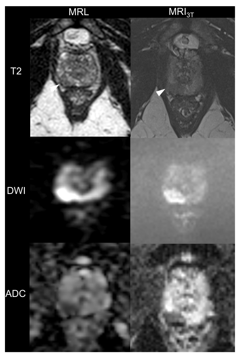
The objective of this study is to conduct a qualitative and a quantitative image quality and lesion evaluation in patients undergoing MR-guided radiation therapy (MRgRT) for prostate cancer on a hybrid magnetic resonance imaging and linear accelerator system (MR-Linac or MRL) at 1.5 Tesla. This prospective study was approved by the institutional review board. A total of 13 consecutive patients with biopsy-confirmed prostate cancer and an indication for MRgRT were included. Prior to radiation therapy, each patient underwent an MR-examination on an MRL and on a standard MRI scanner at 3 Tesla (MRI3T). Three readers (two radiologists and a radiation oncologist) conducted an independent qualitative and quantitative analysis of T2-weighted (T2w) and diffusion-weighted images (DWI). Qualitative outcome measures were as follows: zonal anatomy, capsule demarcation, resolution, visibility of the seminal vesicles, geometric distortion, artifacts, overall image quality, lesion conspicuity, and diagnostic confidence. All ratings were performed on an ordinal 4-point Likert scale. Lesion conspicuity and diagnostic confidence were firstly analyzed only on MRL. Afterwards, these outcome parameters were analyzed in consensus with the MRI3T. Quantitative outcome measures were as follows: anteroposterior and right left diameter of the prostate, lesion size, PI-RADS score (Prostate Imaging-Reporting and Data System) and apparent diffusion coefficient (ADC) of the lesions. Intergroup comparisons were computed using the Wilcoxon-sign rank test and t tests. A post-hoc regression analysis was computed for lesion evaluation. Finally, inter-/intra-reader agreement was analyzed using the Fleiss kappa and intraclass correlation coefficient. For T2w images, the MRL showed good results across all quality criteria (median 3 and 4). Furthermore, there were no significant differences between MRL and MRI3T regarding capsule demarcation or geometric distortion. For the DWI, the MRL performed significantly less than MRI3T across most image quality criteria with a median ranging between 2 and 3. However, there were no significant differences between MRL and MRI3T regarding geometric distortion. In terms of lesion conspicuity and diagnostic confidence, inter-reader agreement was fair for MRL alone (Kappa = 0.42) and good for MRL in consensus with MRI3T (Kappa = 0.708). Thus, lesion conspicuity and diagnostic confidence could be significantly improved when reading MRL images in consensus with MRI3T (Odds ratio: 9- to 11-fold for the T2w images and 5- to 8-fold for the DWI) (p < 0.001). For measures of lesion size, anterior-posterior and right-left prostate diameter, inter-reader and intersequence agreement were excellent (ICC > 0.90) and there were no significant differences between MRL and MRI3T among all three readers. In terms of Prostate Imaging Reporting and Data System (PIRADS) scoring, no significant differences were observed between MRL and MRI3T. Finally, there was a significant positive linear relationship between lesion ADC measurements (r = 0.76, p < 0.01) between the ADC values measured on both systems. In conclusion, image quality for T2w was comparable and diagnostic even without administration of spasmolytic- or contrast agents, while DWI images did not reach diagnostic level and need to be optimized for further exploitation in the setting of MRgRT. Diagnostic confidence and lesion conspicuity were significantly improved by reading MRL in consensus with MRI3T which would be advisable for a safe planning and treatment workflow. Finally, ADC measurements of lesions on both systems were comparable indicating that, lesion ADC as measured on the MRL could be used as a biomarker for evaluation of treatment response, similar to examinations using MRI3T.
Keywords: MR-Linac; PIRADS; adaptive radiotherapy; image guidance; image quality; lesion detection; mpMRI; prostate carcinoma.
Conflict of interest statement
The authors’ institution operates a Linac Unity (Elekta, Sweden). The authors declare no further conflicts of interest. We mention the cooperation with Siemens Healthcare, Philips, Elekta and PTB Braunschweig in another research project (D.W., D.Z., D.T., A.-C.M.). The aforementioned entities had no role in the design of the study; in the collection, analyses, or interpretation of data; in the writing of the manuscript, or in the decision to publish the results.
Figures


References
-
- Kooreman E.S., van Houdt P.J., Nowee M.E., van Pelt V.W., Tijssen R.H., Paulson E.S., Gurney-Champion O.J., Wang J., Koetsveld F., van Buuren L.D., et al. Feasibility and accuracy of quantitative imaging on a 1.5 T MR-linear accelerator. Radiother. Oncol. 2019;133:156–162. doi: 10.1016/j.radonc.2019.01.011. - DOI - PubMed
-
- Hehakaya C., Zyp J.R.V.D.V.V., Lagendijk J.J.W., Grobbee D.E., Verkooijen H.M., Moors E.H.M. Problems and Promises of Introducing the Magnetic Resonance Imaging Linear Accelerator Into Routine Care: The Case of Prostate Cancer. Front. Oncol. 2020;10:1741. doi: 10.3389/fonc.2020.01741. - DOI - PMC - PubMed
Grants and funding
LinkOut - more resources
Full Text Sources
Other Literature Sources
Research Materials
Miscellaneous
