

Defective Oligodendroglial Lineage and Demyelination in Amyotrophic Lateral Sclerosis
- PMID: 33810425
- PMCID: PMC8036314
- DOI: 10.3390/ijms22073426
Defective Oligodendroglial Lineage and Demyelination in Amyotrophic Lateral Sclerosis
Abstract
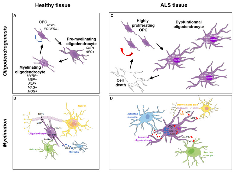
Motor neurons and their axons reaching the skeletal muscle have long been considered as the best characterized targets of the degenerative process observed in amyotrophic lateral sclerosis (ALS). However, the involvement of glial cells was also more recently reported. Although oligodendrocytes have been underestimated for a longer time than other cells, they are presently considered as critically involved in axonal injury and also conversely constitute a target for the toxic effects of the degenerative neurons. In the present review, we highlight the recent advances regarding oligodendroglial cell involvement in the pathogenesis of ALS. First, we present the oligodendroglial cells, the process of myelination, and the tight relationship between axons and myelin. The histological abnormalities observed in ALS and animal models of the disease are described, including myelin defects and oligodendroglial accumulation of pathological protein aggregates. Then, we present data that establish the existence of dysfunctional and degenerating oligodendroglial cells, the chain of events resulting in oligodendrocyte degeneration, and the most recent molecular mechanisms supporting oligodendrocyte death and dysfunction. Finally, we review the arguments in support of the primary versus secondary involvement of oligodendrocytes in the disease and discuss the therapeutic perspectives related to oligodendrocyte implication in ALS pathogenesis.
Keywords: axonal degeneration; demyelination; oligodendrocyte.
Conflict of interest statement
The authors declare no conflict of interest.
Figures
Similar articles
-
Cell-autonomous requirement of TDP-43, an ALS/FTD signature protein, for oligodendrocyte survival and myelination.Proc Natl Acad Sci U S A. 2018 Nov 13;115(46):E10941-E10950. doi: 10.1073/pnas.1809821115. Epub 2018 Oct 29. Proc Natl Acad Sci U S A. 2018. PMID: 30373824 Free PMC article.
-
Abnormal Upregulation of GPR17 Receptor Contributes to Oligodendrocyte Dysfunction in SOD1 G93A Mice.Int J Mol Sci. 2020 Mar 31;21(7):2395. doi: 10.3390/ijms21072395. Int J Mol Sci. 2020. PMID: 32244295 Free PMC article.
-
Myelin degeneration induced by mutant superoxide dismutase 1 accumulation promotes amyotrophic lateral sclerosis.Glia. 2019 Oct;67(10):1910-1921. doi: 10.1002/glia.23669. Epub 2019 Jul 10. Glia. 2019. PMID: 31290185
-
The role of oligodendroglial dysfunction in amyotrophic lateral sclerosis.Neurodegener Dis Manag. 2014;4(3):223-39. doi: 10.2217/nmt.14.21. Neurodegener Dis Manag. 2014. PMID: 25095817 Review.
-
Oligodendroglia: metabolic supporters of neurons.J Clin Invest. 2017 Sep 1;127(9):3271-3280. doi: 10.1172/JCI90610. Epub 2017 Sep 1. J Clin Invest. 2017. PMID: 28862639 Free PMC article. Review.
Cited by
-
Critical analysis of translational potential of rodent models of white matter pathology across a wide spectrum of human diseases.Cell Death Dis. 2025 Jul 31;16(1):580. doi: 10.1038/s41419-025-07893-6. Cell Death Dis. 2025. PMID: 40744926 Free PMC article. Review.
-
lncRNA Sequencing Reveals Neurodegeneration-Associated FUS Mutations Alter Transcriptional Landscape of iPS Cells That Persists in Motor Neurons.Cells. 2023 Oct 16;12(20):2461. doi: 10.3390/cells12202461. Cells. 2023. PMID: 37887305 Free PMC article.
-
The Node of Ranvier as an Interface for Axo-Glial Interactions: Perturbation of Axo-Glial Interactions in Various Neurological Disorders.J Neuroimmune Pharmacol. 2023 Jun;18(1-2):215-234. doi: 10.1007/s11481-023-10072-z. Epub 2023 Jun 7. J Neuroimmune Pharmacol. 2023. PMID: 37285016 Review.
-
Schwann Cells in Neuromuscular Disorders: A Spotlight on Amyotrophic Lateral Sclerosis.Cells. 2025 Jan 3;14(1):47. doi: 10.3390/cells14010047. Cells. 2025. PMID: 39791748 Free PMC article. Review.
-
Icariin prevents methylmercury-induced experimental neurotoxicity: Evidence from cerebrospinal fluid, blood plasma, brain samples, and in-silico investigations.Heliyon. 2024 Jan 6;10(1):e24050. doi: 10.1016/j.heliyon.2024.e24050. eCollection 2024 Jan 15. Heliyon. 2024. PMID: 38226245 Free PMC article.
References
Publication types
MeSH terms
Substances
Grants and funding
LinkOut - more resources
Full Text Sources
Other Literature Sources
Medical
Miscellaneous
