Sodium Fluoride In Vitro Treatment Affects the Expression of Gonadotropin and Steroid Hormone Receptors in Chicken Embryonic Gonads
- PMID: 33810503
- PMCID: PMC8066272
- DOI: 10.3390/ani11040943
Sodium Fluoride In Vitro Treatment Affects the Expression of Gonadotropin and Steroid Hormone Receptors in Chicken Embryonic Gonads
Abstract
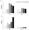
Sodium fluoride (NaF), in addition to preventing dental decay may negatively affect the body. The aim of this study was to examine the effect of a 6 h in vitro treatment of gonads isolated from 14-day-old chicken embryos with NaF at doses of 1.7 (D1), 3.5 (D2), 7.1 (D3), and 14.2 mM (D4). The mRNA expression of luteinizing hormone receptor (LHR), follicle-stimulating hormone receptor (FSHR), estrogen receptors (ESR1 and ESR2), progesterone receptor (PGR), and the immunolocalization of progesterone receptors were examined in the tissue. In the ovary, the expression of FSHR and LHR increased following the NaF treatment. In the case of FSHR the highest stimulatory effect was noticed in the D2 group, while the expression of LHR increased in a dose-dependent manner. A gradual increase in ESR1 and PGR mRNA levels was also observed in the ovary following the NaF treatment, but only up to the D3 dose of NaF. The highest ESR2 level was also found in the D3 group. In the testes, the lowest dose of NaF significantly decreased the expression of FSHR, ESR1, ESR2, and PGR. On the other hand, an increase in PGR expression was observed in the D3 group. The expression of LHR in the testes was not affected by the NaF treatment. Immunohistochemical analysis showed that NaF exposure increased progesterone receptor expression in the ovarian cortex, while it decreased its expression in the testes. These results reveal that NaF may disturb the chicken embryonic development and different mechanisms of this toxicant action exist within the females and males.
Keywords: NaF; chicken embryo; gonads; hormone receptors.
Conflict of interest statement
The authors declare no conflict of interest.
Figures


Similar articles
-
The toxicity mechanism of sodium fluoride on fertility in female rats.Food Chem Toxicol. 2013 Dec;62:566-72. doi: 10.1016/j.fct.2013.09.023. Epub 2013 Sep 23. Food Chem Toxicol. 2013. PMID: 24071475
-
Immunolocalization of GnRHRI, gonadotropin receptors, PGR, and PGRMCI during follicular development in the rabbit ovary.Theriogenology. 2014 May;81(8):1139-47. doi: 10.1016/j.theriogenology.2014.01.043. Epub 2014 Feb 10. Theriogenology. 2014. PMID: 24612788
-
Effect of in vitro sodium fluoride treatment on CAT, SOD and Nrf mRNA expression and immunolocalisation in chicken (Gallus domesticus) embryonic gonads.Theriogenology. 2020 Nov;157:263-275. doi: 10.1016/j.theriogenology.2020.07.020. Epub 2020 Jul 24. Theriogenology. 2020. PMID: 32823022
-
Endometriosis and nuclear receptors.Hum Reprod Update. 2019 Jul 1;25(4):473-485. doi: 10.1093/humupd/dmz005. Hum Reprod Update. 2019. PMID: 30809650 Free PMC article. Review.
-
Gonadotropin receptors and the control of gonadal steroidogenesis: physiology and pathology.Baillieres Clin Endocrinol Metab. 1998 Apr;12(1):35-66. doi: 10.1016/s0950-351x(98)80444-8. Baillieres Clin Endocrinol Metab. 1998. PMID: 9890061 Review.
Cited by
-
Transcriptomic analysis of melatonin on the mechanism of embryonic gonadal development in female Jilin white geese.Poult Sci. 2025 Jan;104(1):104527. doi: 10.1016/j.psj.2024.104527. Epub 2024 Nov 10. Poult Sci. 2025. PMID: 39566171 Free PMC article.
-
Ameliorative Effect of Artemisia absinthium Ethanolic Extract Against Sodium Fluoride Toxicity in Rat Testes: Histological and Physiological Study.Vet Sci. 2025 Apr 15;12(4):371. doi: 10.3390/vetsci12040371. Vet Sci. 2025. PMID: 40284873 Free PMC article.
References
Grants and funding
LinkOut - more resources
Full Text Sources
Other Literature Sources
Research Materials
Miscellaneous

