Review
. 2021 Mar 26;13(7):1538.
doi: 10.3390/cancers13071538.

Mechanisms of Resistance to PI3K Inhibitors in Cancer: Adaptive Responses, Drug Tolerance and Cellular Plasticity

Affiliations
Review

Mechanisms of Resistance to PI3K Inhibitors in Cancer: Adaptive Responses, Drug Tolerance and Cellular Plasticity

Sarah Christine Elisabeth Wright et al. Cancers (Basel). .

Abstract

The phosphatidylinositol-3-kinase (PI3K) pathway plays a central role in the regulation of several signalling cascades which regulate biological processes such as cellular growth, survival, proliferation, motility and angiogenesis. The hyperactivation of this pathway is linked to tumour progression and is one of the most common events in human cancers. Additionally, aberrant activation of the PI3K pathway has been demonstrated to limit the effectiveness of a number of anti-tumour agents paving the way for the development and implementation of PI3K inhibitors in the clinic. However, the overall effectiveness of these compounds has been greatly limited by inadequate target engagement due to reactivation of the pathway by compensatory mechanisms. Herein, we review the common adaptive responses that lead to reactivation of the PI3K pathway, therapy resistance and potential strategies to overcome these mechanisms of resistance. Furthermore, we highlight the potential role in changes in cellular plasticity and PI3K inhibitor resistance.

Keywords: PI3K pathway; PI3K pathway inhibitors; mechanisms of resistance.


Conflict of interest statement

The authors declare no conflict of interest.

Figures

Figure 1
Figure 1
The PI3K and MAPK signalling pathways. Growth factors and other external signalling molecules stimulate the activation of the PI3K and MAPK pathways through receptor tyrosine kinase (RTK) phosphorylation at the cell membrane. Activated RTKs recruit molecules bearing phosphotyrosine-binding (PTB) or Src homology-2 (SH2) domains, such as IRS or p85, respectively. For PI3K enzymes, the binding of the p85 subunit activates the catalytic function of p110, prompting the phosphorylation of phosphatidylinositol 4,5-bisphosphate (PI(4,5)P2) to phosphatidylinositol 3,4,5-trisphosphate (PI(3,4,5)P3). PI(3,4,5)P3, a prominent second messenger in cell signalling, accumulates at the cell membrane attracting molecules with pleckstrin-homology (PH) domains, primarily phosphoinositide-dependent protein kinase 1 (PDK1) and the AKT serine/threonine kinase family (protein kinase B/PKB). PDK1 partially activates AKT at threonine 308 through phosphorylation, with full activation enabled by serine 473 phosphorylation via the mammalian target of rapamycin (mTOR) complex 2 (mTORC2). AKT targets various proteins in order to alter major signalling pathways within the cell. These include prosurvival pathways (BIM, BAX, BAD, BCL-2, MDM2 and FoxO1), cell cycle progression and glucose metabolism (p27, GSK3 and AS160), and cellular proliferation and protein synthesis (TSC2). Another prominent effector targeted downstream in the PI3K pathway is mTOR complex 1 (mTORC1), which has been shown to regulate cellular growth and metabolism, amongst other processes. In particular, mTORC1 targets ribosomal protein S6 kinase (S6K) and eukaryotic initiation factor 4E-binding protein 1 (4E-BP1), with the former directly effecting eukaryotic initiation factor 4E (eIF4E) and subsequent translation of cell cycle regulators.
Figure 2
Figure 2
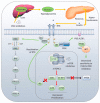
Insulin-mediated feedback loops following PI3K/mTOR inhibition. Following treatment with PI3K inhibitors, the liver breaks down stored glycogen releasing glucose into the bloodstream. The increased levels of glucose (hyperglycaemia) are detected by the pancreas, and in an effort to overcome these high levels of glucose, large amounts of insulin are released (hyperinsulinemia). This substantial release of insulin is sufficient to partially reactivate the insulin receptor which re-instates both IRS and GBR2 activity. What results is an increase in both PI3K and MAPK pathway activation, limiting the therapeutic effects of PI3K inhibitors. mTOR1 inhibitors, such as rapamycin, block downstream translation by downregulating S6K1 and 4E-BP1. In turn, this de-represses the S6K1 substrate IRS1, which acts as an intermediary between insulin receptor and the PI3K complex. The recruitment of PI3K to the active receptor enhances both MAPK and downstream PI3K signalling. Thus, limiting the overall sensitivity of mTOR inhibition in these tumours. Downregulation of mTOR activity either through AKT inhibition or direct mTOR inhibition blocks 4E-BP1-mediated translation of PTEN. This enhances the pool of PIP3 in cells resulting in the sustained activation of AKT.
Figure 3
Figure 3
Adaptive and epigenetic-driven mechanisms of resistance to PI3K/AKT inhibition. A number of adaptive mechanisms regulating PI3K inhibitor sensitivity have been described. The most notable of which is the upregulation receptor tyrosine kinases (RTKs) following PI3K/AKT inhibition. Members of the FOXO family of transcription factors are direct substrates of AKT with phosphorylation limiting their nuclear localisation. Upon AKT downregulation FOXO is drawn into the nucleus where it is recruited to binding sites which upregulate a number of receptor tyrosine kinases including HER2 and HER3. This upregulation of these RTKs results in the enhanced activity of both MAPK and PI3K signalling pathways. Similarly, in ER+ breast cancers AKT phosphorylates the methyl-transferase KMT2D inhibiting the methyl-transferase activity of the enzyme. Upon AKT inhibition KMT2D activity is restored priming the recruitment of the transcription factors FOXA1, PBX1 and ER onto ER binding sites enhancing ER-dependent gene transcription and limiting the PI3K therapeutic effects. SGK3 is an ER transcriptional target. PIK3CA mutant tumours displaying low levels of pAKT can circumvent this by activating SGK3 and downstream mTOR signalling limiting the sensitivity of PI3K inhibitors.

Similar articles

Cited by

References

    1. Cantley L.C., Whitman M., Chahwala S., Fleischman L., Kaplan D.R., Schaffhausen B.S., Roberts T.M. Oncogenes and phosphatidylinositol turnover. Ann. N. Y. Acad. Sci. 1986;488:481–490. doi: 10.1111/j.1749-6632.1986.tb46580.x. - DOI - PubMed
    1. Thorpe L.M., Yuzugullu H., Zhao J.J. PI3K in cancer: Divergent roles of isoforms, modes of activation and therapeutic targeting. Nat. Rev. Cancer. 2015;15:7–24. doi: 10.1038/nrc3860. - DOI - PMC - PubMed
    1. Engelman J.A., Luo J., Cantley L.C. The evolution of phosphatidylinositol 3-kinases as regulators of growth and metabolism. Nat. Rev. Genet. 2006;7:606–619. doi: 10.1038/nrg1879. - DOI - PubMed
    1. Wong K.K., Engelman J.A., Cantley L.C. Targeting the PI3K signaling pathway in cancer. Curr. Opin. Genet. Dev. 2009;20:87–90. doi: 10.1016/j.gde.2009.11.002. - DOI - PMC - PubMed
    1. Vanhaesebroeck B., Leevers S.J., Panayotou G., Waterfield M.D. Phosphoinositide 3-kinases: A conserved family of signal transducers. Trends Biochem. Sci. 1997;22:267–272. doi: 10.1016/S0968-0004(97)01061-X. - DOI - PubMed

LinkOut - more resources