Review
. 2021 Apr;11(4):971-994.
doi: 10.1158/2159-8290.CD-21-0010.

Metastasis-Initiating Cells and Ecosystems

Affiliations
Review

Metastasis-Initiating Cells and Ecosystems

Joan Massagué et al. Cancer Discov. 2021 Apr.

Abstract

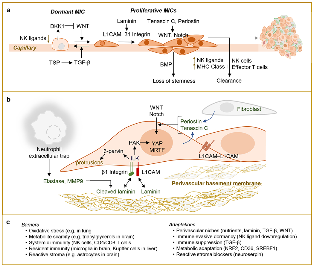
Metastasis is initiated and sustained through therapy by cancer cells with stem-like and immune-evasive properties, termed metastasis-initiating cells (MIC). Recent progress suggests that MICs result from the adoption of a normal regenerative progenitor phenotype by malignant cells, a phenotype with intrinsic programs to survive the stresses of the metastatic process, undergo epithelial-mesenchymal transitions, enter slow-cycling states for dormancy, evade immune surveillance, establish supportive interactions with organ-specific niches, and co-opt systemic factors for growth and recurrence after therapy. Mechanistic understanding of the molecular mediators of MIC phenotypes and host tissue ecosystems could yield cancer therapeutics to improve patient outcomes. SIGNIFICANCE: Understanding the origins, traits, and vulnerabilities of progenitor cancer cells with the capacity to initiate metastasis in distant organs, and the host microenvironments that support the ability of these cells to evade immune surveillance and regenerate the tumor, is critical for developing strategies to improve the prevention and treatment of advanced cancer. Leveraging recent progress in our understanding of the metastatic process, here we review the nature of MICs and their ecosystems and offer a perspective on how this knowledge is informing innovative treatments of metastatic cancers.


Conflict of interest statement

Conflict of Interest Disclosure: JM owns stock of Scholar Rock Inc. One or both authors are inventors in the following patents and patent applications: US Patent 7829066 (JM); EU Patent 2831593 (JM); EU Patent 3047039 (JM). US Patent 10413522 (JM); Patent Application PCT/US2016/062880 (JM); and, Patent Application PCT/US2017/045145 (JM and KG).

Figures

Figure 1.
Figure 1.. Phases of Metastasis.
a. Metastasis proceeds through three distinct phases of dissemination, dormancy and colonization. Metastasis Initiating Cells (MICs) disseminate from the primary tumor and seed multiple organs, where they enter a subclinical state of dormancy. During dormancy, MICs may shuttle between quiescent and proliferative states, with proliferative cells being continually cleared by niche-specific or systemic immune defenses. MICs that acquire immune evasive and organ-specific growth adaptations are able to exit dormancy and generate clinically evident macrometastatic colonies. During dissemination and dormancy, MICs are in a dynamic equilibrium with host immunity, while failure of immune surveillance results in metastatic outbreaks and organ colonization. b. The three phases of metastasis can overlap with the growth of primary tumors and may co-exist in the same individual until removal of the primary tumor (Stage I-III). The latter two phases continue to co-exist during adjuvant therapy, with the eventual dominance of the colonization phase resulting in macrometastatic relapse (in cases that were originally diagnosed as Stage I-III), or in patients diagnosed with de novo Stage IV cancers.
Figure 2.
Figure 2.. Metastatic dissemination.
The most common and best characterized route of tumor dissemination is via the blood circulation (a-c). a. During hematogenous dissemination, cancer cells at the invasion front of primary tumors that undergo epithelial-mesenchymal transitions (EMT) lead intravasation into neoangiogenic capillaries in the tumor, and thus access the venous circulation for dissemination to multiple organs. Cancer cells may intravasate individually or in clusters during collective migration. Invasion and intravasation involve remodeling of the tumor extracellular matrix (ECM) and may be facilitated by tumor-resident fibroblasts and macrophages. b. In the circulation, cancer cells must rapidly adapt to overcome biomechanical, redox and immunological threats. Clustering of circulating tumor cells (CTCs) may enable paracrine growth factor signaling that promotes the niche-independent survival of disseminated epithelial cells. CTC clusters are enriched in stem-like cancer cells and can include other cell types including platelets and neutrophils, which in turn can protect CTCs from immune attack by secreting immunosuppressive factors. c. CTCs become trapped in the capillary beds of multiple organs and migrate into the organ parenchyma to seed nascent metastasis. Fenestrated capillary beds in the liver and bone marrow can facilitate extravasation. Non-tumor cells with CTC clusters, or in the parenchyma, including platelets and monocytes, can facilitate endothelial permeability and transmigration by secreting endothelial disjunction factors. Alternatively, CTCs can secrete factors that induce endothelial necroptosis. d. Beyond hematogenous dissemination, cancer cells originating in certain primary tumors can also reach distant organs via alternative routes.
Figure 3.
Figure 3.. Metastatic dormancy and outbreak.
a. Metastasis initiating cells (MICs) seeding distant organs enter into a variable period of dormancy, when then cannot be detected by clinical imaging technologies. Clinical dormancy reflects an equilibrium incorporating cell-intrinsic growth arrest, and stochastic MIC proliferation events, which are countered by elimination of proliferating cells. Secreted factors in the perivascular microenvironment may either inhibit MIC proliferation (e.g. TGF-β), or promote it (e.g. WNT), which in turn can be countered by MIC secretion of WNT inhibitors (e.g. DKK1). MICs spread on the abluminal surface of capillaries by adhering to the perivascular basement membrane, which provides proliferative inputs. However, stromal factors can induce proliferating MICs to differentiate and lose stemness properties (e.g. BMP). In contrast to quiescent MICs, proliferating MICs upregulate cell surface expression of NK ligands and MHC Class I molecules, facilitating their detection and destruction by NK cells and adaptive immune cells. b. Exit from dormancy requires close interactions between MICs and extracellular matrix (ECM). MICs induce innate immune responses that include the recruitment of neutrophils and the formation of extracellular web-like chromatin decondensates harboring ECM remodeling enzymes, termed neutrophil extracellular traps (NETs). NET-dependent cleavage of laminin, a key component of the ECM, activates β1-integrin signaling in MICs. MICs can also activate signaling through integrin-like kinase (ILK) by upregulating L1CAM, a cell adhesion molecule that binds laminin on extracellular basement membranes, including that of blood capillaries. Integrin/ILK signaling enables cell stretching via the formation of actin-dependent protrusions, which in turn enables mechanosignaling to activate YAP-dependent transcriptional output and proliferation. Homophilic L1CAM interactions as cancer cells multiply can extend this signaling. Additional cues from the ECM, including the stem cell niche ECM components tenascin C and periostin, can potentiate growth factor signaling and enable MIC stemness and proliferation. c. Outgrowth of dormant metastasis might be prevented by bolstering barriers to MIC survival and proliferation or targeting the phenotypic adaptations required for escape from dormancy.
Figure 4.
Figure 4.. Acquisition of MIC phenotypes.
a. MICs arise from primary tumors but must acquire distinct phenotypic traits in order to successfully disseminate, seed and colonize distant organs. MICs adopt key phenotypic traits of regenerative progenitors that respond to tissue injury, including phenotypic plasticity, the ability to restore heterogeneous and morphologically complex epithelial structures upon disruption of tissue structure, and the ability to evade killing by immune cells. In addition, MICs must acquire organ-specific adaptations that enable colonization of distinct microenvironments. b. Models of metastasis evolution. In the parallel evolution model, cancer cells disseminating from the primary tumor early during cancer progression seed distant organs and evolve genetically, independently from the primary tumor. Thus, mutations in the primary and metastatic tumors would be expected to be different. In contrast, in linear evolution models, MICs disseminate late during tumor evolution from the primary tumor, and thus are closely clonally related to the primary tumor. Linear evolution can be gradual, in which case tumors may consist of multiple distinct subclones with similar fitness and individual metastases may be derived from distinct subclones, or punctuated, in which case a dominant subclone arising in the primary tumor rapidly outcompetes and overtakes the entire population and seeds all metastases. Dash arrows represent the length of metastasis latency time periods.
Figure 5.
Figure 5.. Model of the regenerative progenitor origin of MICs.
a. In the intestine, proliferative LGR5+ crypt base cells maintain intestinal homeostasis. Injury disrupts intercellular adherens junctions and induces the emergence of an L1CAM+ regenerative progenitor phenotype, which is required for epithelial repair, regeneration of heterogeneous cell types, and resolution of the wound. b. Oncogenic driver mutations in homeostatic stem and progenitor cells create tumor initiating cells (or “cancer stem cells”, CSCs), capable of adenoma formation within an intact epithelial niche. Additional mutations give rise to an invasive carcinoma, which further breaches the integrity of the epithelium and triggers the emergence of an L1CAM+ regenerative progenitor state in cancer cells. As regenerative progenitors, these malignant cells have the capacity to survive the loss of the epithelial niche during tumor dissemination, are competent to enter quiescence, evade immune surveillance and therapy, and upon the receipt of favorable growth signals, regenerate heterogeneous tumors in metastatic sites and after therapy.
Figure 6.
Figure 6.. Metastasis as a systemic disease with organ specific features.
a. A recap of organ-specific determinants of metastatic colonization. b. Systemic determinants of metastasis. During metastasis, cancer cells from primary tumors disseminate throughout the body via the blood and lymphatic circulations, among other routes. Tumor cells establish a dynamic equilibrium with both systemic immunity and organ-specific immune infiltrates. Systemic factors can modulate both primary and metastatic tumor growth as well as the anti-tumor immune responses. While the catalog of systemic factors that can modulate metastasis is incomplete, current evidence implicates circulating chemokines and cytokines and signals transmitted via nerves and hormones in controlling tumor growth and metastasis. In addition, intratumoral and gut microbiomes can influence cancer progression either through direct interactions with cancer cells and immune cells, or indirectly, via metabolites released into the systemic circulation. The dynamic equilibrium between tumors, metastases and their niches is influenced by systemic factors that impact MIC growth and therapy resistance.

References

    1. Lambert AW, Pattabiraman DR, Weinberg RA. Emerging Biological Principles of Metastasis. Cell 2017;168(4):670–91 doi 10.1016/j.cell.2016.11.037. - DOI - PMC - PubMed
    1. Massagué J, Obenauf AC. Metastatic colonization by circulating tumour cells. Nature 2016;529(7586):298–306 doi 10.1038/nature17038. - DOI - PMC - PubMed
    1. Oskarsson T, Batlle E, Massagué J. Metastatic stem cells: sources, niches, and vital pathways. Cell Stem Cell 2014;14(3):306–21 doi 10.1016/j.stem.2014.02.002. - DOI - PMC - PubMed
    1. Batlle E, Clevers H. Cancer stem cells revisited. Nat Med 2017;23(10):1124–34 doi 10.1038/nm.4409. - DOI - PubMed
    1. Celià-Terrassa T, Kang Y. Metastatic niche functions and therapeutic opportunities. Nature Cell Biology 2018;20(8):868–77 doi 10.1038/s41556-018-0145-9. - DOI - PubMed

Publication types