. 2021 Sep;86(3):1299-1313.
doi: 10.1002/mrm.28784. Epub 2021 Apr 3.

Specific absorption rate and temperature in neonate models resulting from exposure to a 7T head coil

Affiliations

Specific absorption rate and temperature in neonate models resulting from exposure to a 7T head coil

Shaihan J Malik et al. Magn Reson Med. 2021 Sep.

Abstract

Purpose: To investigate safe limits for neonatal imaging using a 7T head coil, including both specific absorption rate (SAR) and temperature predictions.

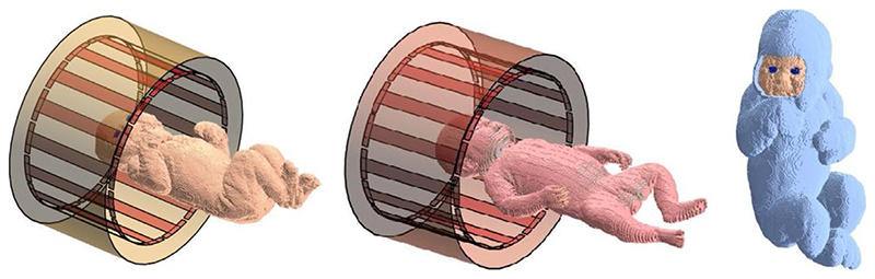
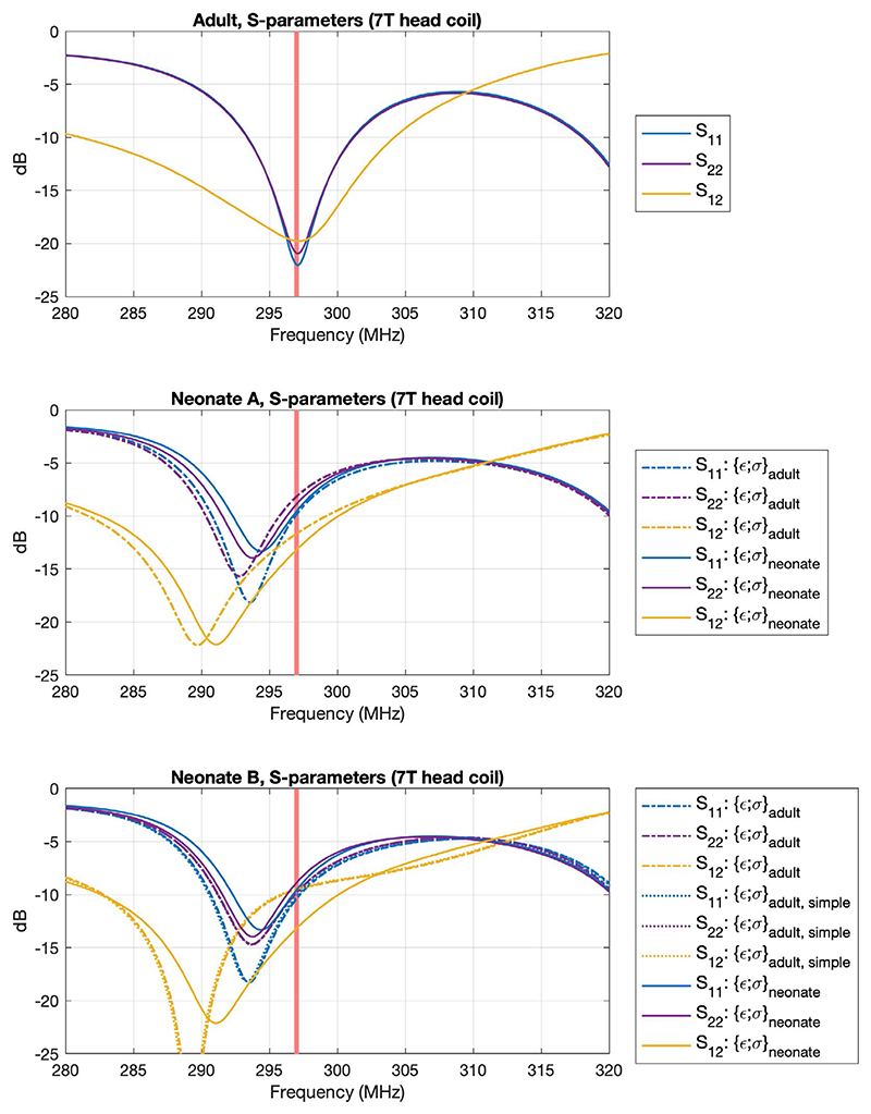
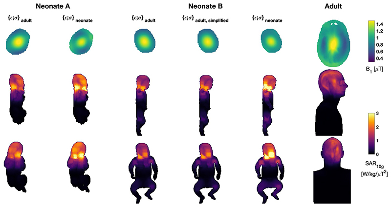
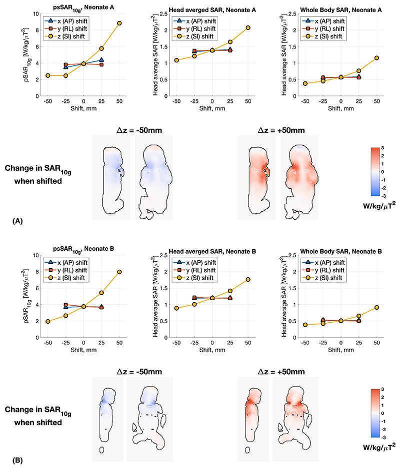
Methods: Head-centered neonate models were simulated using finite-difference time domain-based electromagnetic and thermal solvers. The effects of higher water content of neonatal tissues compared with adults, position shifts, and thermal insulation were also considered. An adult model was simulated for comparison.

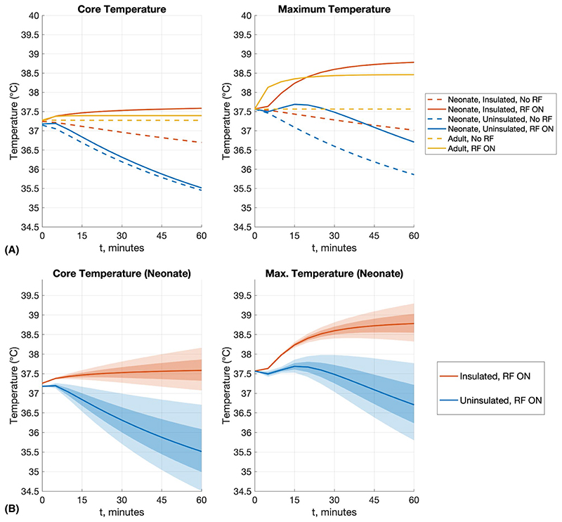
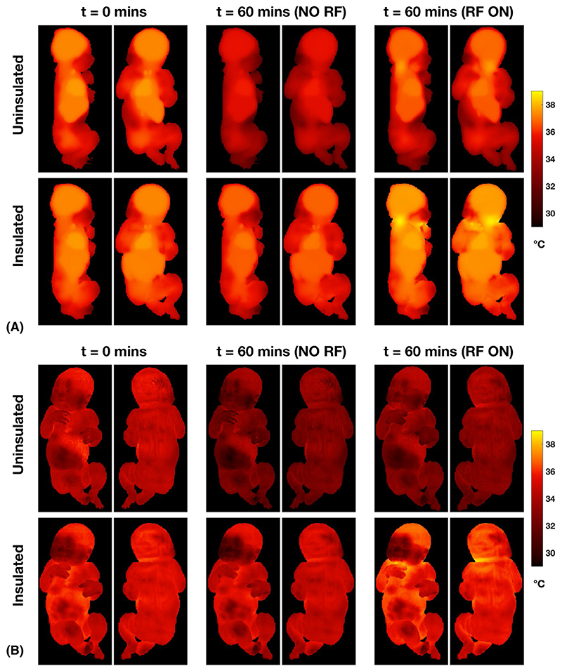
Results: Maximum and average SAR are both elevated in the neonate when compared with an adult model. When normalized to B1+ , the SAR experienced by a neonate is greater than an adult by approximately a factor of 2; when normalized to net forward power (forward-reflected), this increases to a factor of 2.5-3.0; and when normalized to absorbed power, approximately a factor of 4. Use of age-adjusted dielectric properties significantly increases the predicted SAR, compared with using adult tissue properties for the neonates. Thermal simulations predict that change in core temperature/maximum temperature remain compliant with International Electrotechnical Commission limits when a thermally insulated neonate is exposed at the SAR limit for up to an hour.

Conclusion: This study of two neonate models cannot quantify the variability expected within a larger population. Likewise, the use of age-adjusted dielectric properties have a significant effect, but while their use is well motivated by literature, there is uncertainty in the true dielectric properties of neonatal tissue. Nevertheless, the main finding is that unlike at lower field strengths, operational limits for 7T neonatal MRI using an adult head coil should be more conservative than limits for use on adults.

Keywords: 7T MRI; RF safety; neonatal imaging.


Figures

Figure 1
Figure 1
Neonates within transmit coil and shield. Left: Neonate A. Center: Neonate B. The large shield representing the bore of the scanner is not shown. Right: Neonate A with insulating “blanket”
Figure 2
Figure 2
S-parameters of the simulated birdcage coil (driven at two ports) for each of the voxel models simulated. The coil was tuned and matched at 297 MHz (indicated by red line) using the adult voxel model, and generally good matching can be seen for this model (Snn≤ 20 dB). We observe some shifting of the resonance frequency when loaded with neonate models, but still obtain acceptable return loss. Neonates were simulated both with adult ({ε;σ}adult) and neonate-appropriate ({ε;σ}neonate) dielectric properties. Neonate B was also simulated with a simplified tissue segmentation ({ε;σ}adult,simple) (details in text)
Figure 3
Figure 3
The B1+ and 10g specific absorption rate (SAR10g) for all neonate models and Duke. The neonate models were simulated using both adult and neonate specific (age-adjusted) dielectric properties. Neonate B was also simulated using a simplified tissue segmentation to match the lower number of separate tissue classes in model A. Models are all depicted at the same spatial scale. Top row: B1+ distributions in central axial plane for mean B1+ = 1 μT; this slice is the one used to normalize the SAR distributions also reported in Table 1. Middle/bottom rows: SAR10g distributions as maximum projections in sagittal/coronal views, respectively
Figure 4
Figure 4
Summary of changes in simulated SAR when the neonate models are shifted inside the coil (A, neonate A; B, neonate B; both cases using neonatal dielectric properties). In each case, the SAR values are normalized by the average B1+ within the same anatomic slice (shown in Figure 3) (ie, the slice position is shifted as the model is shifted). Inset images show the representative slices highlighting changes in SAR10g with respect to no spatial shift. Abbreviations: AP, anterior–posterior; RL, right–left; SI, superior–inferior
Figure 5
Figure 5
Simulated B1+ distributions and homogeneity measure (coefficient of variation) for cylinders of different diameter within the 7T head coil. Cylinders had the same dielectric properties as muscle. B1+ distributions are shown at the same relative scales (ie, between maximum and minimum values) to highlight the homogeneity of the pattern rather than the absolute value. As expected, the homogeneity worsens as the diameter increases; however, it remains within a similar range between the diameters of 80 mm and 180 mm
Figure 6
Figure 6
A, Summary temperature results for neonate A (neonatal properties) with and without thermal insulation (“blanket”), and the Duke adult model for comparison. Simulations were run for 60 minutes before this, to establish thermal equilibrium (details in text). For all scenarios, the simulations were run such that head-average SAR = 3.2 W/kg, as this was the most limiting in all cases. B, Because the heat transfer coefficient (h) for neonates is not well understood, simulations were repeated by changing h by ±10% (darker shading) and ±20% (lighter shading)
Figure 7
Figure 7. Temperature distributions for neonate A with and without thermal insulation (blanket).
A, Maximum intensity projections. B, Surface temperature distributions. The starting time (t = 0) was reached after 60 minutes of simulation to reach a stable starting condition. Temperature distributions after a further 60 minutes without (middle columns) or with (right columns) RF exposure are shown. Without thermal insulation or RF exposure, the neonate cools significantly. When insulated and with 60 minutes of RF exposure, higher local temperatures are reached in the posterior part of the neck, and the face/hand where these are close together, but do not exceed 39°C

References

    1. Kelly CJ, Hughes EJ, Rutherford MA, Counsell SJ. Advances in neonatal MRI of the brain: from research to practice. Arch Dis Child— Educ Pract Ed. 2019;104:106–110. - PubMed
    1. Harris AD, Singer HS, Horska A, et al. GABA and glutamate in children with primary complex motor stereotypies: an 1H-MRS study at 7T. Am J Neuroradiol. 2016;37:552–557. - PMC - PubMed
    1. De Vita E, Bainbridge A, Cheong JLY, et al. Magnetic resonance imaging of neonatal encephalopathy at 4.7 Tesla: initial experiences. Pediatrics. 2006;118:e1812–e1821. - PubMed
    1. Food and Drug Administration. Guidance for Industry and Food and Drug Administration Staff— Criteria for Significant Risk Investigations of Magnetic Resonance Diagnostic Devices. 2014. [Accessed March 22, 2021]. https://www.fda.gov/regulatory-information/search-fda-guidance-documents... .
    1. International Electrotechnical Commission. IEC 60601-2-33: Medical Electrical Equipment—Particular Requirements for the Safety of Magnetic Resonance Equipment for Medical Diagnosis. 2015. [Accessed March 22, 2021]. https://webstore.iec.ch/publication/22705 .

Publication types