. 2021 Aug;175(4):920-930.
doi: 10.1002/ajpa.24272. Epub 2021 Apr 3.

Novel strategies for the characterization of cancellous bone morphology: Virtual isolation and analysis

Affiliations

Novel strategies for the characterization of cancellous bone morphology: Virtual isolation and analysis

Alessio Veneziano et al. Am J Phys Anthropol. 2021 Aug.

Abstract

Objectives: The advent of micro-computed tomography (μCT) made cancellous bone more accessible than ever before. Nevertheless, the characterization of cancellous bone is made difficult by its inherent complexity and the difficulties in defining homology across datasets. Here we propose novel virtual methodological approaches to overcome those issues and complement existing methods.

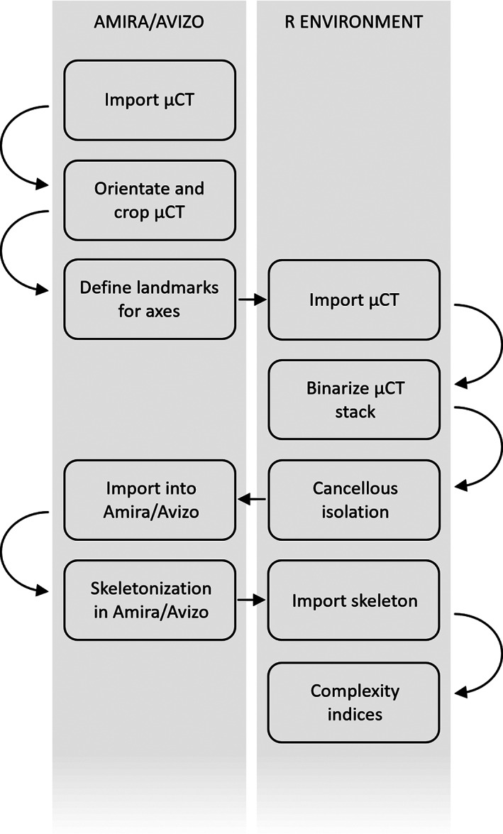
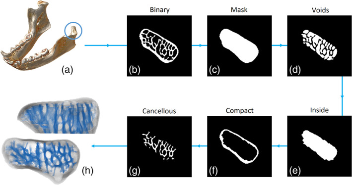
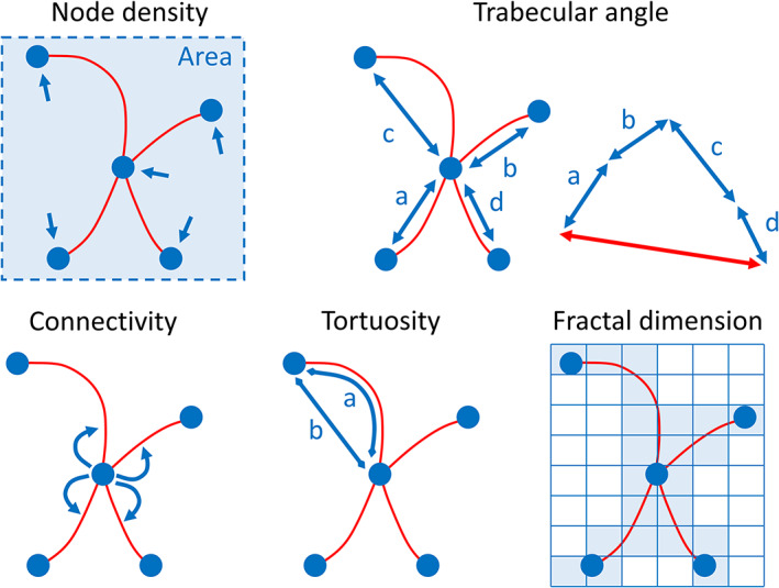
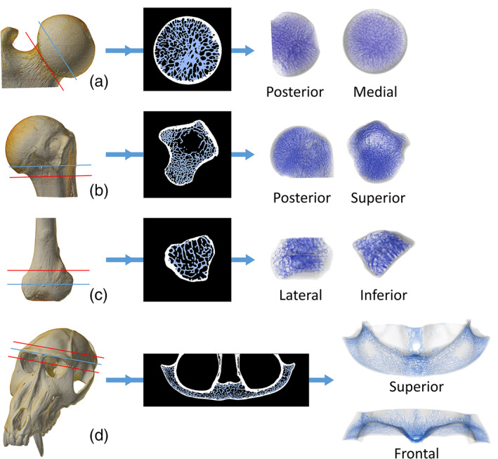
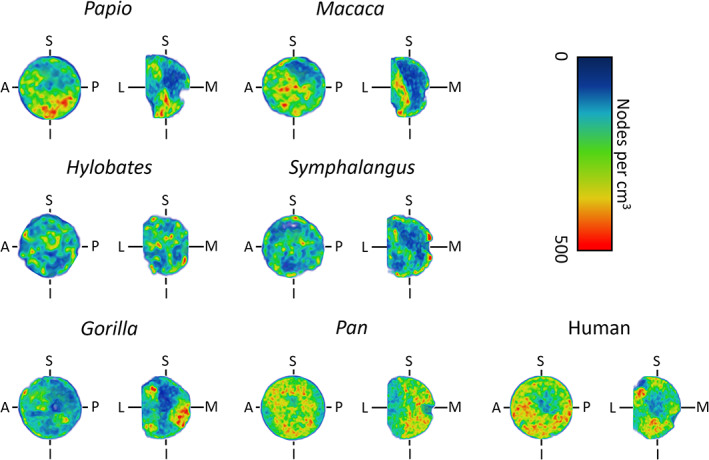
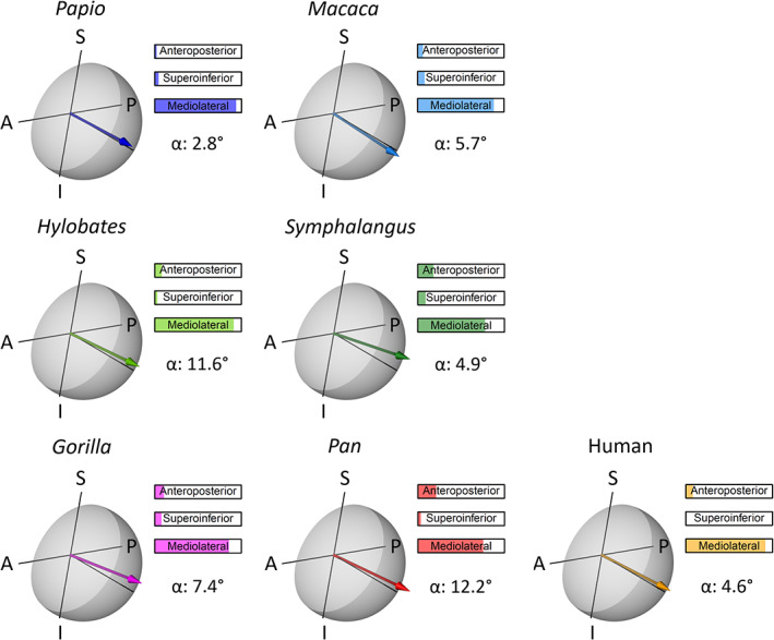
Materials and methods: We present a protocol for the isolation of the whole cancellous region within a μCT scanned bone. This method overcomes the subsampling issues and allows studying cancellous bone as a single unit. We test the protocol on a set of primate bones. In addition, we describe a set of morphological indices calculated on the topological skeleton of the cancellous bone: node density, node connectivity, trabecular angle, trabecular tortuosity, and fractal dimension. The usage of the indices is shown on a small comparative sample of primate femoral heads.

Results: The isolation protocol proves reliable in isolating cancellous structures from several different bones, regardless of their shape. The indices seem to detect some functional differences, although further testing on comparative samples is needed to clarify their potential for the study of cancellous architecture.

Conclusions: The approaches presented overcome some of the difficulties of trabecular bone studies. The methods presented here represent an alternative or supporting method to the existing tools available to address the biomechanics of cancellous bone.

Keywords: bone complexity; bone segmentation; primates; skeletonization; trabecular architecture.


Figures

FIGURE 1
FIGURE 1
The workflow of the methodological approach introduced in this work. Actions are divided based on the environment where they are performed (R or Amira/Avizo)
FIGURE 2
FIGURE 2
The protocol for the semi‐automatic isolation of cancellous bone shown on the mandibular condyle of Hylobates lar. The region of interest (a) is cropped out of the μCT scan and the volume is binarised (b). The binarised image enters the first step of the protocol. Multiple dilations and erosions fill the empty spaces surrounding the cancellous bone, creating a mask (c) of the whole bone region. By subtracting the binary image from the mask (c minus b), the voids are highlighted (d). The voids undergo multiple dilations and erosions, returning the area occupied by voids and cancellous bone (e), which is within the compact bone. By subtracting the inside area from the mask (c minus e), the compact bone is isolated (f). The cancellous bone (g) is then obtained by subtracting the compact bone and the voids from the mask (c minus d minus f). The operation is performed on single μCT slices stacked to obtain a 3D result (h, superior and frontal views of the mandibular condyle)
FIGURE 3
FIGURE 3
Graphical intuition of the indices measured on the topological skeleton of cancellous bone. For ease of visualization, the indices are shown for a 2D topological skeleton. Node density is represented by the number of nodes per unit area and it is calculated using a kernel density approximation over a discretized space. The trabecular angle (degrees) is measured between a reference axis (not shown) and the unitary resultant (red, double‐headed arrow) of all trabecular directions (blue, double‐headed arrows) obtained by vector sum. Connectivity is the mean number of branches connected to non‐terminal nodes. Tortuosity is the ratio between the arc length of a branch and the linear distance between its starting and ending nodes (a/b). Fractal dimension is an index of complexity measured on the coordinates of the skeleton using the box‐counting algorithm. In this approach, discrete regular grids of decreasing cell size are superimposed over the cancellous skeleton and the number of cells occupied by the skeleton are counted for each grid. Fractal dimension is the slope of the line fitting the number of cells that overlap the skeleton versus the inverse of the cell size
FIGURE 4
FIGURE 4
Semi‐automatic isolation of cancellous bone in the femoral head of Symphalangus syndactylus (a), the proximal humerus of Alouatta caraya (b), the distal fibula of Cercopithecus albogularis (c) and the brow ridge of Mandrillus sphynx (d). The 3D μCT scan is cut (red line) to limit the cancellous isolation to a region of interest. The results are here shown on a single 2D slice (indicated by the blue line on the 3D scan) and on the full 3D μCT stack (the cutting planes used to isolate the 3D regions of interest is shown in red)
FIGURE 5
FIGURE 5
Node density of the femoral head, measured using a kernel density approximation over a regular 3D grid. It is expressed as the number of nodes of the skeletonized cancellous bone per cm3. The node density is here shown for a small sample of primates over the coronal (L‐M‐S‐I) and para‐sagittal (A‐P‐S‐I) planes. The density increases from blue to red. (A, anterior; P, posterior; S, superior; I, inferior; L, lateral; M, medial)
FIGURE 6
FIGURE 6
Trabecular angle of the femoral head calculated as the 3D angle between the mediolateral axis and the resultant of all trabecular directions. Trabecular directions are measured on the branches of the skeletonized cancellous bone. The trabecular angle is here shown for a small sample of primates on a transparent model of the femoral head. The mediolateral axis is the line perpendicular to the A‐P‐S‐I plane (para‐sagittal plane). The anteroposterior, supero‐inferior, and mediolateral percentage contributions to the angle are reported. The arrow point is for easing visualization only and does not indicate a verse. (A, anterior; P, posterior; S, superior; I, inferior)

References

    1. Andreassen, T. T., & Oxlund, H. (2001). The effects of growth hormone on compact and cancellous bone. Journal of Musculoskeletal and Neuronal Interactions, 2(1), 49–58. - PubMed
    1. Annadhason, A. (2012). Methods of fractal dimension computation. International Journal of Computer Science and Information Technology & Security., 2(1), 166–169.
    1. Bishop, P. J., Clemente, C. J., Hocknull, S. A., Barrett, R. S., & Lloyd, D. G. (2017). The effects of cracks on the quantification of the cancellous bone fabric tensor in fossil and archaeological specimens: A simulation study. Journal of Anatomy, 230(3), 461–470. 10.1111/joa.12569 - DOI - PMC - PubMed
    1. Bishop, P. J., Hocknull, S. A., Clemente, C. J., Hutchinson, J. R., Farke, A. A., Barrett, R. S., & Lloyd, D. G. (2018). Cancellous bone and theropod dinosaur locomotion. Part III—Inferring posture and locomotor biomechanics in extinct theropods, and its evolution on the line to birds. PeerJ, 6, e5777. 10.7717/peerj.5777 - DOI - PMC - PubMed
    1. Carter, D. R., Orr, T. E., & Fyhrie, D. P. (1989). Relationships between loading history and femoral cancellous bone architecture. Journal of Biomechanics, 22(3), 231–244. 10.1016/0021-9290 - DOI - PubMed

Publication types

LinkOut - more resources