Review
. 2021 Jul 12;56(13):1861-1874.
doi: 10.1016/j.devcel.2021.03.020. Epub 2021 Apr 2.

Extracellular vesicles: Critical players during cell migration

Affiliations
Review

Extracellular vesicles: Critical players during cell migration

Bong Hwan Sung et al. Dev Cell. .

Abstract

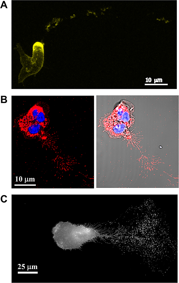
Cell migration is essential for the development and maintenance of multicellular organisms, contributing to embryogenesis, wound healing, immune response, and other critical processes. It is also involved in the pathogenesis of many diseases, including immune deficiency disorders and cancer metastasis. Recently, extracellular vesicles (EVs) have been shown to play important roles in cell migration. Here, we review recent studies describing the functions of EVs in multiple aspects of cell motility, including directional sensing, cell adhesion, extracellular matrix (ECM) degradation, and leader-follower behavior. We also discuss the role of EVs in migration during development and disease and the utility of imaging tools for studying the role of EVs in cell migration.

Keywords: adhesion; cell migration; cell motility; chemotaxis; exosomes; extracellular vesicles; live imaging; microvesicles; migrasomes.


Conflict of interest statement

Declaration of interests The authors declare no competing interests.

Figures

Figure 1.
Figure 1.. EV secretion promotes the directional migration of cells.
Cells migrating toward a gradient of soluble chemoattractants secrete extracellular vesicles (EVs) promoting their directional migration. Microvesicles (MVs) bud directly from the plasma membrane. Released MVs carry matrix metalloproteases (MMPs) degrading extracellular matrix to promote cancer cell invasion. Also, tissue transglutaminse 2 (TG2)-carrying MVs activate focal adhesion kinase (FAK) to promote cell contractility. Another type of large EV called a migrasome is formed at the tips or intersections of retraction fibers and enriched with tetraspanins (TSPANs) 4 and 7. Chemokines associated with migrasomes, such as CXCL12, released from leader cells (cyan cell) can promote chemotaxis of follower cells (orange cell) in a paracrine manner. Exosome functions in cell migration are depicted as: A. Multivesicular bodies (MVBs) are formed by inward budding of late endosomal membranes. Endosomal sorting complex required for transport (ESCRT) machinery, neutral sphingomyelinase 2 (nSMASE2), tetraspanins (TSPANs), and Alix-Syndecan-Syntenin complex are all molecules known to drive MVB biogenesis by inducing formation of intraluminal vesicles (ILV). These ILVs are secreted as exosomes after fusion of MVBs with the plasma membrane. B. Exosomes secreted at the leading edge of a migrating cell promote adhesion formation by binding integrin receptors at the cell membrane through fibronectin presented on the surface of the exosomes. C. Cortical branched actin filaments stabilized by cortactin enhance MVB docking at invadopodial protrusions. The secreted exosomes promote invadopodia formation and carry proteinases such as MT1-MMP that degrade ECM and promote cancer cell invasion. D. In Dictyostelium, exosomes carry the adenylyl cyclase ACA, which synthesizes the chemoattractant cAMP in the exosomal lumen and is secreted through specific transporters. The secreted exosomal attractants promote both autocrine and paracrine migration. Created with BioRender.com
Figure 2.
Figure 2.. Cells leave trails of EVs behind during migration.
A. Fluorescence maximum intensity projection of Dictyostelium discoideum aca cells expressing ACA-YFP and chemotaxing towards cAMP. Note the trails of ACA-YFP-positive vesicles left from the rear of the migrating cell. Reproduced under the term of the Creative Commons CC BY-NC-SA 3.0 license (Kriebel et al., 2008). Copyright © 2008, Rockefeller University Press. B. Confocal images of primary human neutrophils migrating towards fMLF, fixed and stained using an antibody against CD63 (red) and the nuclear dye Hoechst (blue). The image shows CD63-positive vesicles left behind the migrating neutrophil. C. Fluorescence image of HT1080 fibrosarcoma cells stably expressing pHluo_M153R-CD63. Note the exosome trails left behind the cell as it migrates toward the left side of the image. Reproduced under the term of the Creative Commons CC BY 4.0 license (Sung et al., 2020). Copyright © 2020, Springer Nature.
Figure 3.
Figure 3.. Cells exhibit pathfinding behavior on EV trails.
A. Left: Fluorescence maximum intensity images taken from a time lapse recording of Dictyostelium discoideum aca cells expressing ACA-YFP moving towards an aggregate of cells. Cell outline traces depicting the relative position of 4 cells is presented as a time evolution of outlines from lighter to darker colors for each cell. The secreted ACA-YFP-containing vesicles are depicted in gray. Right: Plot showing the correlation between the distance of a cell to its nearest vesicle and the angle θ between the cell direction of motion and the direction vector to the nearest vesicle. In this context, the closer a cell is to a vesicle, the higher the chemotactic response of the cells, which is measured by smaller angles of deviation. Reproduced under the term of the Creative Commons CC BY-NC-SA 4.0 license (Kriebel et al., 2018). Copyright © 2018, Rockefeller University Press. B. Left: Fluorescence time series images of HT1080 fibrosarcoma cells co-expressing pHluo_M153R-CD63 (exosome label) and mCherry-CAAX (plasma membrane label), showing leader-follower cell migration behavior in 3D collagen gels. Exosome trails (arrowheads) deposited behind migrating leader cells promote directional migration of follower cells (arrows). Right: Scatter plot with median and quartile range showing pathfinding index quantitated by the cosine value of the angle θ between the cell direction of motion and the nearest exosome trail. Note that migration towards exosome trails yields positive cosine θ values, with perfect migration along an exosome trail yielding a cosine θ = 1, whereas migration away from exosome trails yields negative cosine θ values, with a maximum possible value of −1. Reproduced under the term of the Creative Commons CC BY 4.0 license (Sung et al., 2020). Copyright © 2020, Springer Nature.

References

    1. Afonso PV, Janka-Junttila M, Lee YJ, McCann CP, Oliver CM, Aamer KA, Losert W, Cicerone MT, and Parent CA (2012). LTB4 is a signal-relay molecule during neutrophil chemotaxis. Developmental cell 22, 1079–1091. - PMC - PubMed
    1. Altei WF, Pachane BC, Dos Santos PK, Ribeiro LNM, Sung BH, Weaver AM, and Selistre-de-Araújo HS (2020). Inhibition of αvβ3 integrin impairs adhesion and uptake of tumor-derived small extracellular vesicles. Cell Commun Signal 18, 158–158. - PMC - PubMed
    1. Antonyak MA, Li B, Boroughs LK, Johnson JL, Druso JE, Bryant KL, Holowka DA, and Cerione RA (2011). Cancer cell-derived microvesicles induce transformation by transferring tissue transglutaminase and fibronectin to recipient cells. Proceedings of the National Academy of Sciences 108, 4852. - PMC - PubMed
    1. Awojoodu AO, Keegan PM, Lane AR, Zhang Y, Lynch KR, Platt MO, and Botchwey EA (2014). Acid sphingomyelinase is activated in sickle cell erythrocytes and contributes to inflammatory microparticle generation in SCD. Blood 124, 1941–1950. - PMC - PubMed
    1. Beer KB, Rivas-Castillo J, Kuhn K, Fazeli G, Karmann B, Nance JF, Stigloher C, and Wehman AM (2018). Extracellular vesicle budding is inhibited by redundant regulators of TAT-5 flippase localization and phospholipid asymmetry. Proceedings of the National Academy of Sciences 115, E1127. - PMC - PubMed

Publication types