. 2021 Apr 6;35(1):108959.
doi: 10.1016/j.celrep.2021.108959. Epub 2021 Mar 23.

Drug repurposing screens reveal cell-type-specific entry pathways and FDA-approved drugs active against SARS-Cov-2

Affiliations

Drug repurposing screens reveal cell-type-specific entry pathways and FDA-approved drugs active against SARS-Cov-2

Mark Dittmar et al. Cell Rep. .

Abstract

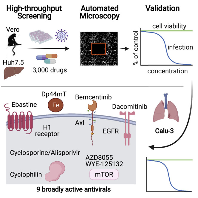
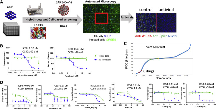
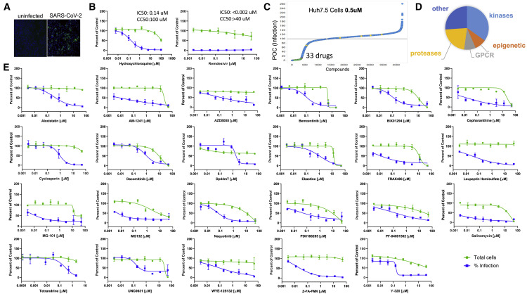
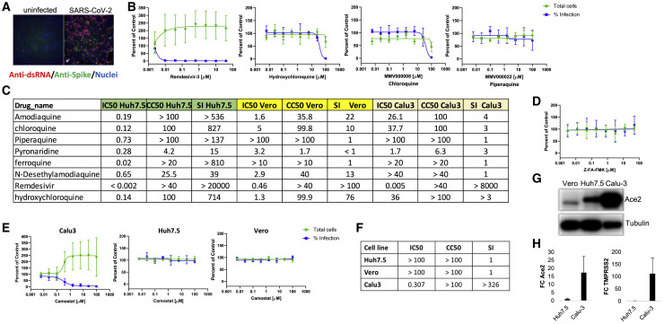
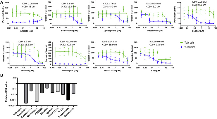
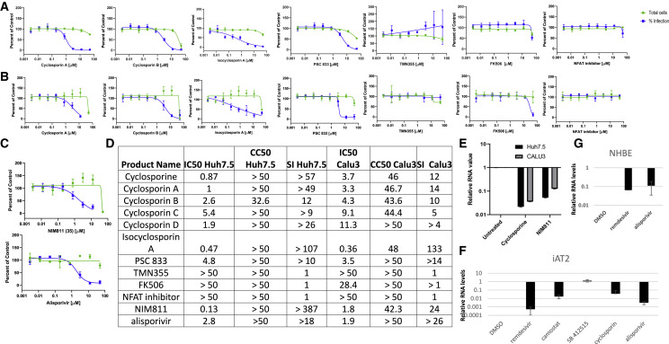
There is an urgent need for antivirals to treat the newly emerged severe acute respiratory syndrome coronavirus 2 (SARS-CoV-2). To identify new candidates, we screen a repurposing library of ∼3,000 drugs. Screening in Vero cells finds few antivirals, while screening in human Huh7.5 cells validates 23 diverse antiviral drugs. Extending our studies to lung epithelial cells, we find that there are major differences in drug sensitivity and entry pathways used by SARS-CoV-2 in these cells. Entry in lung epithelial Calu-3 cells is pH independent and requires TMPRSS2, while entry in Vero and Huh7.5 cells requires low pH and triggering by acid-dependent endosomal proteases. Moreover, we find nine drugs are antiviral in respiratory cells, seven of which have been used in humans, and three are US Food and Drug Administration (FDA) approved, including cyclosporine. We find that the antiviral activity of cyclosporine is targeting Cyclophilin rather than calcineurin, revealing essential host targets that have the potential for rapid clinical implementation.

Keywords: HTS; SARS2; TMPRSS2; antiviral; coronavirus; cyclophilin; cyclosporin; drugs; entry; screening.


Conflict of interest statement

Declaration of interests The authors declare no competing interests.

Figures

None
Graphical abstract
Figure 1
Figure 1
High-throughput screening in Vero cells to identify antivirals against SARS-CoV-2 (A) Schematic of the screening strategy. Vero cells were plated in 384-well plates, drugs were added, and the cells were infected with SARS-CoV-2 (MOI = 1). 30 hpi cells were stained for viral infection (dsRNA, Spike) and imaged using automated microscopy to define cell number and percent infection. Antivirals show little impact on cell number and block viral infection. (B) Dose-response analysis of Vero cells treated with hydroxychloroquine or remdesivir. Each data point represents the average of two independent experiments ± SD. (C) Percent of control (POC) for percentage of infection of the Vero drug screen performed at 1 μM. Six drugs had >60% reduction in infection with >80% cell viability. (D) Dose-response analysis of six candidates identified in the screen. Each data point represents the average of ≥2 independent experiments ± SD.
Figure 2
Figure 2
High-throughput screening in human Huh7.5 cells to identify antivirals against SARS-CoV-2 (A) Huh7.5 cells were infected with SARS-CoV-2 (MOI = 1) and 30 hpi processed for microscopy. (B) Dose-response analysis of Huh7.5 cells treated with hydroxychloroquine or remdesivir. Data represent the average of three independent experiments ± SD. (C) POC for percentage of infection of the Huh7.5 drug screen performed at 0.5 μM. 33 drugs had >60% reduction in infection with >80% cell viability. (D) Distribution of 23 validated antivirals by drug target class. (E) Dose-response analysis of the candidates with a selectivity index (SI) > 3 identified in the screen. Data represent the average of three independent experiments ± SD.
Figure 3
Figure 3
Cell-type-specific dependencies of entry inhibitors (A) Calu-3 human lung epithelial cells were infected with SARS-CoV-2 (MOI = 0.5) and processed for microscopy 48 hpi. (B) Dose-response analysis of Calu-3 cells treated with quinolines or remdesivir. Data represent the average of four independent experiments ± SD. (C) IC50, CC50, and SI for Vero, Huh7.5, and Calu-3 cells treated with a panel of quinolines or remdesivir. Data represent the average of four independent experiments ± SD. (D) Dose-response analysis of Calu-3 cells treated with the cathepsin inhibitor Z-FA-FMK. Data represent the average of four independent experiments ± SD. (E) Dose response analysis of Calu-3, Vero, and Huh7.5 cells treated with camostat. Data represent the average of ≥2 independent experiments ± SD. (F) IC50, CC50, and SI for camostat across cell types. (G) Immunoblot of Vero, Huh7.5, and Calu-3 cells probed for Ace2 and tubulin as a loading control. Representative blot is shown. (H) qRT-PCR of Ace2 or TMPRSS2 comparing Huh7.5 and Calu-3 cells. Data represent the mean ± SEM for ≥2 independent experiments.
Figure 4
Figure 4
Validation of antiviral activity of nine candidates in Calu-3 cells (A) Dose-response analysis of Huh7.5 candidate antivirals in Calu-3 cells with a SI > 3. Data represent the average of ≥4 independent experiments ± SD. (B) qRT-PCR analysis of nine candidate antivirals in Calu-3 cells. Data represent the mean ± SEM for ≥2 independent experiments.
Figure 5
Figure 5
Cyclosporine is antiviral against SARS-CoV-2 independent of calcineurin (A) Dose-response analysis of Huh7.5 cells treated with a panel of cyclosporins and related drugs. Data represent the average of ≥2 independent experiments ± SD. (B) Dose-response analysis of Calu-3 cells treated with a panel of cyclosporins and related drugs. Data represent the average of five independent experiments ± SD. (C) Dose-response analysis of Calu3 cells treated with cyclophilin-selective drugs NIM811 and alisporivir. Data represent the average of five independent experiments ± SD. (D) Table of IC50s, CC50s, and SI of Huh7.5 cells and Calu-3 cells treated with the indicated drugs. (E) qRT-PCR analysis of viral replication in Huh7.5 and Calu-3 cells treated with the indicated drugs. (F) qRT-PCR analysis of viral replication in iAT2 cells treated with the indicated drugs. (G) qRT-PCR of NHBE cells treated with the indicated drug. For (E)–(G), data represent the mean ± SEM for ≥2 independent experiments.

Similar articles

Cited by

References

    1. Ashburn T.T., Thor K.B. Drug repositioning: identifying and developing new uses for existing drugs. Nat. Rev. Drug Discov. 2004;3:673–683. - PubMed
    1. Bailly C. Cepharanthine: An update of its mode of action, pharmacological properties and medical applications. Phytomedicine. 2019;62:152956. - PMC - PubMed
    1. Beigel J.H., Tomashek K.M., Dodd L.E., Mehta A.K., Zingman B.S., Kalil A.C., Hohmann E., Chu H.Y., Luetkemeyer A., Kline S., et al. Remdesivir for the Treatment of Covid-19 - Preliminary Report. Reply. N. Engl. J. Med. 2020;383:994. - PubMed
    1. Belouzard S., Chu V.C., Whittaker G.R. Activation of the SARS coronavirus spike protein via sequential proteolytic cleavage at two distinct sites. Proc. Natl. Acad. Sci. USA. 2009;106:5871–5876. - PMC - PubMed
    1. Blanco-Melo D., Nilsson-Payant B.E., Liu W.-C., Uhl S., Hoagland D., Møller R., Jordan T.X., Oishi K., Panis M., Sachs D., et al. Imbalanced Host Response to SARS-CoV-2 Drives Development of COVID-19. Cell. 2020;181:1036–1045.e9. - PMC - PubMed

Publication types