Review
. 2021 Jul:68:101335.
doi: 10.1016/j.arr.2021.101335. Epub 2021 Mar 31.

Reactive astrocytes: The nexus of pathological and clinical hallmarks of Alzheimer's disease

Affiliations
Review

Reactive astrocytes: The nexus of pathological and clinical hallmarks of Alzheimer's disease

Brittani R Price et al. Ageing Res Rev. 2021 Jul.

Abstract

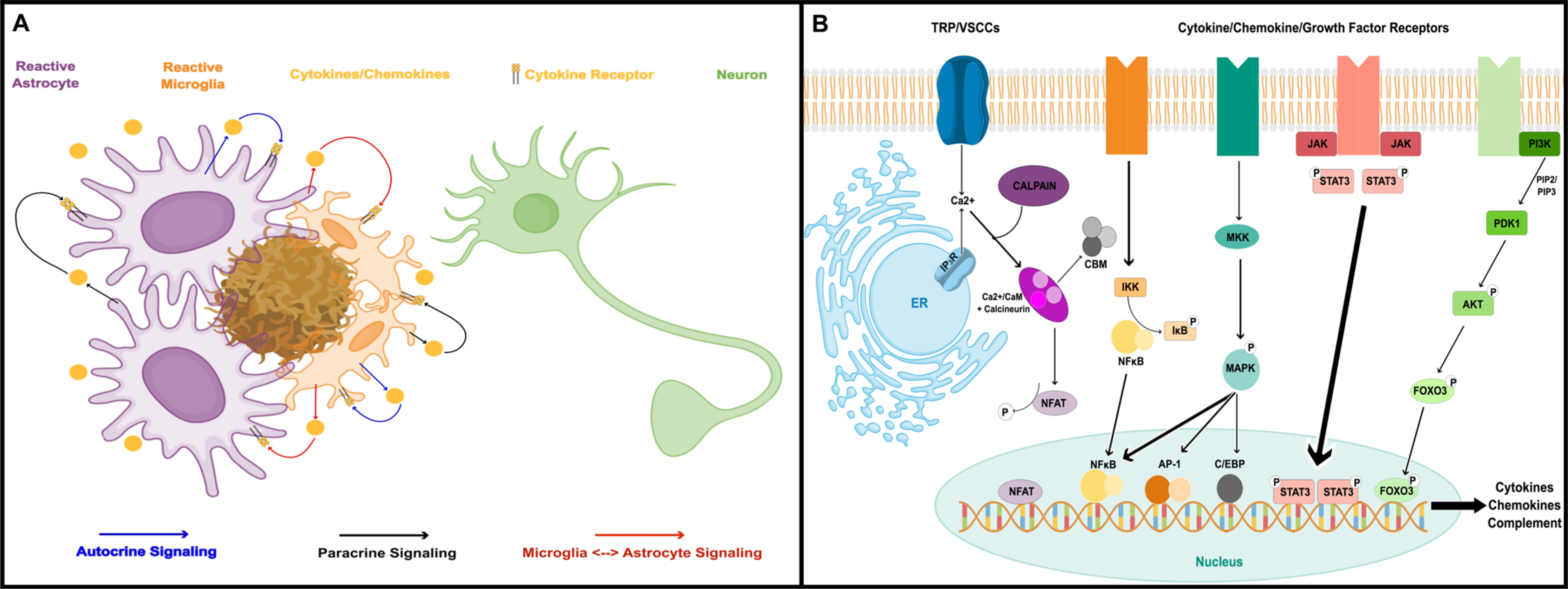
Astrocyte reactivity is a hallmark of neuroinflammation that arises with Alzheimer's disease (AD) and nearly every other neurodegenerative condition. While astrocytes certainly contribute to classic inflammatory processes (e.g. cytokine release, waste clearance, and tissue repair), newly emerging technologies for measuring and targeting cell specific activities in the brain have uncovered essential roles for astrocytes in synapse function, brain metabolism, neurovascular coupling, and sleep/wake patterns. In this review, we use a holistic approach to incorporate, and expand upon, classic neuroinflammatory concepts to consider how astrocyte dysfunction/reactivity modulates multiple pathological and clinical hallmarks of AD. Our ever-evolving understanding of astrocyte signaling in neurodegeneration is not only revealing new drug targets and treatments for dementia but is suggesting we reimagine AD pathophysiological mechanisms.

Keywords: Alzheimer’s disease; Astrocytes; Dementia; Neurodegeneration; Neuroinflammation; Reactive astrocytes.


Figures

Fig. 1.
Fig. 1.. Reactive astrocytes are a key component to neuroinflammation in AD.
A) Cartoon showing astrocytes and microglia surrounding a parenchymal amyloid deposit. Both cell types respond to amyloid (and other extracellular factors) with morphologic and biochemical changes including the release of numerous cytokines, chemokines, and other inflammatory factors which can maintain and/or propagate glial reactivity. Neuroinflammatory processes resulting from chronic glial activation can have many deleterious (and sometimes beneficial) effects on neurons. B) Inflammatory factors trigger astrocyte reactivity through a number of transcriptional pathways involving NFκB, MAPK, Jak/Stat, and/or FOXO3. Concurrent Ca2+ signaling and/or Ca2+ dysregulation leads to the activation of the calcineurin/NFAT pathway, which further shapes astrocyte reactivity through extensive interactions with other transcription factors and second/third messenger systems. These pathways, in turn, regulate the production of numerous cytokines and chemokines involved in triggering and maintaining glial reactivity.
Fig. 2.
Fig. 2.. Astrocytes regulate synaptic transmission, synapse stability, and synapse removal.
Pre- and postsynaptic neuronal compartments are cradled by specialized astrocytic processes that express numerous factors that directly shape synapse/structure and function. Astrocyte secreted factors (ASF), including thrombospondin (TSP), hevin, and sparc help anchor pre- and postsynaptic cell adhesion molecules together. This arrangement not only aligns and stabilizes presynaptic terminals with dendritic spines, but it also helps to cluster important synaptic machinery, including postsynaptic density constituents and neurotransmitter receptors, to active zones. Reactive astrocytes in AD brain tissue may release less of the pro-synaptogenetic factors thrombospondin and hevin, relative to sparc (which opposes hevin-mediated synaptogenesis), leading to a net loss of synapses. Astrocytes are also a major source for complement C3, which is released by reactive astrocytes and binds to C3 receptors (C3R) on “weakened” pre- and postsynaptic elements, leading to microglial-mediated phagocytosis. Increased C3 levels arising from reactive astrocytes have been shown to contribute to abnormal synapse loss in mouse models of amyloid pathology. Finally, astrocytes express several different types of glutamate transporters (EAATs 1 and 2, aka Glast and Glt-1) that help terminate synaptic glutamate signaling and prevent hyperactivation of extrasynaptic glutamate receptors. The downregulation of EAAT2/Glt-1 levels and/or function in reactive astrocytes is thought to be a primary mechanism for excitotoxic neuronal degeneration during AD.
Fig. 3.
Fig. 3.. Astrocytic metabolic pathways are essential for meeting the energy demands of neurons.
Astrocytes are the major source of lactate in the brain, which appears to be the preferred energy substrate of neurons. Three intertwined sources drive lactate production in astrocytes: glycogen, glucose uptake through GLUT1 transporters, and glutamate uptake through EAATs. Glucose taken up from blood vessels, or derived from the breakdown of glycogen, is converted to pyruvate and subsequently lactate, which is ultimately released to nearby neurons. Glutamate uptake during neuronal activity helps create an electrochemical gradient (with nearby Na+/K+ exchangers) that facilitates both glucose uptake via GLUT1 and the conversion of glucose to lactate. Glutamate uptake via astrocytic EAATs is also recycled back to neurons in the form of glutamine. Inhibition of astrocyte glycogen metabolism at any of these steps has been shown to be detrimental to both acute neural function and for the extended processes of LTP and memory formation, which are disrupted in many rodent models of AD-like pathology.
Fig. 4.
Fig. 4.. Astrocytes modulate neuronal excitability through potassium spatial buffering.
Neuronal excitability relies on inward Na+ and outward K+ fluxes during action potentials. A) Schematic demonstrating the individual phases that comprise a single action potential. Once an action potential is initiated, voltage-gated Na+ channels in the membrane open to allow an influx of Na+ ions. The influx of Na+ further depolarizes the neuronal membrane, in turn opening additional voltage-gated Na+ channels. Once the peak membrane potential is reached, the neuronal membrane begins to repolarize by inactivating voltage-gated Na+ channels and opening voltage-gated K+ channels. The efflux of K+ ions from the neuron results in a decrease in the membrane potential towards the neuron’s resting voltage. B) Through Kir4.1 channels (shown in pink) and the Cx43-containing gap junctions (shown in teal), astrocytes are able to take up excess extracellular K+ and transfer it either into the circulation via the astrocytic end-feet (indicated by the black arrows) or to an area of the brain lacking K+ via their gap junctions (indicated by the red arrows). The removal of K+ ions from the extracellular space following an action potential is critical in order for the neuronal membrane to adequately repolarize and reset channel function for the next action potential to occur. A single action potential can increase the extracellular K+ concentration by as much as 1 mM under normal conditions and ≥10–12 mM under pathologic conditions. Even the relatively small elevations in extracellular K+ observed during physiologic neuronal activity depolarize the neuronal membrane, thereby increasing the probability of action potential propagation. Thus, impaired K+ buffering can lead to hyperexcitability and subsequent excitotoxicity. Murine models of AD manifest hyperexcitability, with some models also exhibiting evident epileptiform and seizure activity. Moreover, early onset hyperexcitability is a well known feature of human AD.
Fig. 5.
Fig. 5.. Waste products are cleared from the brain by a process that requires astrocytes.
Under physiological conditions, AQP4 channels (shown in navy blue) are polarized to astrocytic endfeet and support rapid water movement between the periarterial space and astroglial syncytium. This anatomic arrangement facilitates the convective bulk flow of CSF from the periarterial space across the astrocytic endfeet and into the interstitial space, where it mixes with interstitial fluid (ISF) and waste products such as Aβ (shown in brown). Waste products and excess fluids are then driven toward the perivenous space and ultimately cleared from the brain through the meningeal lymphatic vessels. Altered AQP4 localization has been described in aged brains, whereas loss of perivascular AQP4 has been demonstrated in human AD brains and is associated with increased levels of Aβ and tau pathology. It should be noted that while the astrocytic arbors appear to overlap in this figure, in reality their arbors occupy distinct fields with little to no overlap.
Fig. 6.
Fig. 6.. Mechanisms underlying astrocyte-mediated vascular responses.
From an anatomical standpoint, astrocytes are perfectly positioned to bi-directionally communicate information between neurons and blood vessels. In order to meet the metabolic needs of active neurons, increased neuronal activity induces a rapid vasodilatory response and consequent spatiotemporally restricted delivery of glucose and oxygen. A) Glutamate released from presynaptic neurons acts on astrocytic metabotropic glutamate receptors (mGluR5) resulting in increased intracellular Ca2+. B) PLA2 is activated in response to rises in intracellular Ca2+ concentrations, leading to the generation of arachidonic acid (AA) and its subsequent conversion to either prostaglandin E2 (PGE2) via COX enzymes or to epoxyeicosatrinoic acids (EETs) by CYP2C11 enzymes. Both PGE2 and EETs act on vascular smooth muscle cells to dilate vessels. Increases in intracellular Ca2+ also engage the Ca2+-dependent K+ channel BK (shown in yellow) on the astrocyte endfoot plasma membrane. Activation of BK results in the efflux of K+ into the extracellular space where it is taken up by vascular smooth muscle cells via Kir2.1 or Kir2.2. Like PGE2 and EETs, K+ also induces vasodilation. C) Conversely, in response to high pO2, AA is released from astrocytes and converted into 20-HETE in the vascular smooth muscle cells. The combination of low extracellular adenosine levels and 20-HETE leads to an elevation in smooth muscle cell free Ca2+ and subsequent arteriolar constriction. Thus, neurovascular coupling, which ensures that the brain has a proportionally matched cerebral blood flow in response to local neuronal activity, is largely mediated by astrocytic Ca2+ signaling. Both BBB and NVU breakdown are evident in AD and may impair neurovascular coupling by preventing astrocytes from relaying signals between the vasculature and neuronal circuitry, creating a mismatch between neuronal activity and the provision of oxygen and glucose required to meet metabolic demands.
Fig. 7.
Fig. 7.
Astrocyte reactivity is a primary nexus for the cerebrovascular and neuronal pathologies that arise with AD. The major functional phenotypes associated with astrocyte reactivity include neuroinflammation, impaired glutamate/potassium homeostasis, hypometabolism, and loss of endfoot integrity, all of which are extensively intertwined. Neuroinflammatory pathways directly affect nearby glial cells, neurons, and neurovascular elements (orange arrows). Glutamate and potassium dysregulation directly affect astrocyte metabolism, synapse function, and vascular endothelial cells (green arrows). Breakdown of astrocyte endfoot processes, lead to the loss of BBB integrity, Ca2+ dysregulation, and impaired glymphatic clearance. Impaired astrocyte metabolism directly erodes neuronal viability and glymphatic clearance of interstitial toxins, including Aβ (blue arrows), which, in turn, create an inhospitable environment for neurons marked by elevated neuroinflammation and excitotoxicity (purple arrow).

References

    1. Abdul HM, Sama MA, Furman JL, Mathis DM, Beckett TL, Weidner AM, Patel ES, Baig I, Murphy MP, LeVine H 3rd, Kraner SD, Norris CM, 2009. Cognitive decline in Alzheimer’s disease is associated with selective changes in calcineurin/NFAT signaling. J. Neurosci 29 (41), 12957–12969. - PMC - PubMed
    1. Afridi R, Kim JH, Rahman MH, Suk K, 2020. Metabolic regulation of glial phenotypes: implications in neuron-glia interactions and neurological disorders. Front. Cell. Neurosci 14, 20. - PMC - PubMed
    1. Allaman I, Gavillet M, Belanger M, Laroche T, Viertl D, Lashuel HA, Magistretti PJ, 2010. Amyloid-beta aggregates cause alterations of astrocytic metabolic phenotype: impact on neuronal viability. J. Neurosci 30 (9), 3326–3338. - PMC - PubMed
    1. Allen NJ, Eroglu C, 2017. Cell biology of astrocyte-synapse interactions. Neuron 96 (3), 697–708. - PMC - PubMed
    1. Allweis C, Landau T, Abeles M, Magnes J, 1966. The oxidation of uniformly labelled albumin-bound palmitic acid to CO2 by the perfused cat brain. J. Neurochem 13 (9), 795–804. - PubMed

Publication types