

PAR-4/Ca2+-calpain pathway activation stimulates platelet-derived microparticles in hyperglycemic type 2 diabetes
- PMID: 33812377
- PMCID: PMC8019350
- DOI: 10.1186/s12933-021-01267-w
PAR-4/Ca2+-calpain pathway activation stimulates platelet-derived microparticles in hyperglycemic type 2 diabetes
Abstract
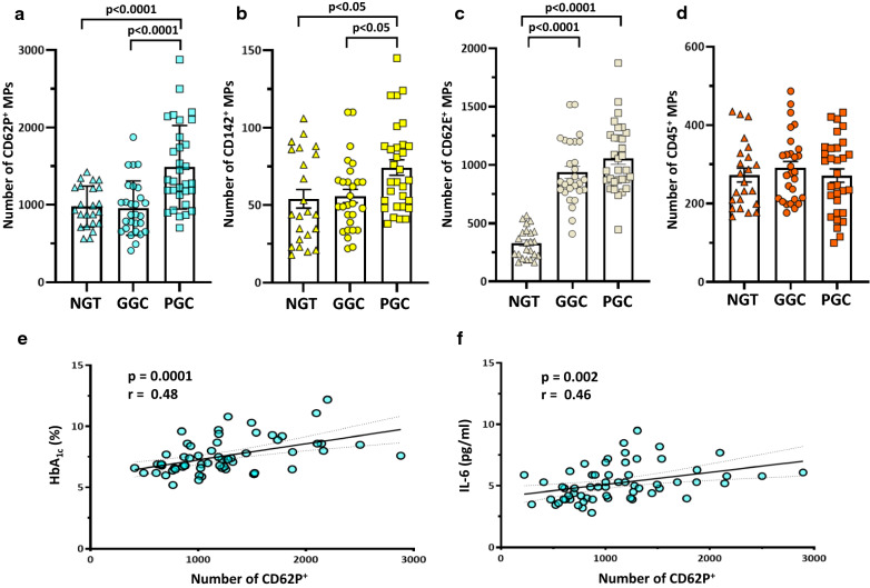
Background: Patients with type 2 diabetes (T2DM) have a prothrombotic state that needs to be fully clarified; microparticles (MPs) have emerged as mediators and markers of this condition. Thus, we investigate, in vivo, in T2DM either with good (HbA1c ≤ 7.0%; GGC) or poor (HbA1c > 7.0%; PGC) glycemic control, the circulating levels of MPs, and in vitro, the molecular pathways involved in the release of MPs from platelets (PMP) and tested their pro-inflammatory effects on THP-1 transformed macrophages.
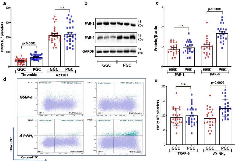
Methods: In 59 T2DM, and 23 control subjects with normal glucose tolerance (NGT), circulating levels of CD62E+, CD62P+, CD142+, CD45+ MPs were determined by flow cytometry, while plasma levels of ICAM-1, VCAM-1, IL-6 by ELISA. In vitro, PMP release and activation of isolated platelets from GGC and PGC were investigated, along with their effect on IL-6 secretion in THP-1 transformed macrophages.
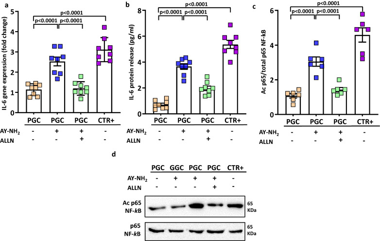
Results: We found that MPs CD62P+ (PMP) and CD142+ (tissue factor-bearing MP) were significantly higher in PGC T2DM than GGC T2DM and NGT. Among MPs, PMP were also correlated with HbA1c and IL-6. In vitro, we showed that acute thrombin exposure stimulated a significantly higher PMP release in PGC T2DM than GGC T2DM through a more robust activation of PAR-4 receptor than PAR-1 receptor. Treatment with PAR-4 agonist induced an increased release of PMP in PGC with a Ca2+-calpain dependent mechanism since this effect was blunted by calpain inhibitor. Finally, the uptake of PMP derived from PAR-4 treated PGC platelets into THP-1 transformed macrophages promoted a marked increase of IL-6 release compared to PMP derived from GGC through the activation of the NF-kB pathway.
Conclusions: These results identify PAR-4 as a mediator of platelet activation, microparticle release, and inflammation, in poorly controlled T2DM.
Keywords: Extracellular vesicles; Glycated hemoglobin; NF-kB; Platelet activation; THP-1 transformed macrophages.
Conflict of interest statement
AA received research grants, lecture or advisory board fees from Merck Sharp & Dome, AstraZeneca, Novartis, Boeringher-Ingelheim, Sanofi, Mediolanum, Janssen, Novo Nordisk, Lilly, Servier, and Takeda, Neopharmed. PS received research grants, lecture or consultant or travel fees from CSL Behring, Stago, Werfen, Uniqure.
Figures


Similar articles
-
Circulating levels and characterization of microparticles in patients with different degrees of glucose tolerance.Cardiovasc Diabetol. 2017 Sep 19;16(1):118. doi: 10.1186/s12933-017-0600-0. Cardiovasc Diabetol. 2017. PMID: 28927403 Free PMC article.
-
Platelet-derived calpain cleaves the endothelial protease-activated receptor 1 to induce vascular inflammation in diabetes.Basic Res Cardiol. 2020 Dec 1;115(6):75. doi: 10.1007/s00395-020-00833-9. Basic Res Cardiol. 2020. PMID: 33258989 Free PMC article.
-
Endothelial microparticle-associated protein disulfide isomerase increases platelet activation in diabetic coronary heart disease.Aging (Albany NY). 2021 Jul 20;13(14):18718-18739. doi: 10.18632/aging.203316. Epub 2021 Jul 20. Aging (Albany NY). 2021. PMID: 34285139 Free PMC article.
-
Identification of a Distinct Platelet Phenotype in the Elderly: ADP Hypersensitivity Coexists With Platelet PAR (Protease-Activated Receptor)-1 and PAR-4-Mediated Thrombin Resistance.Arterioscler Thromb Vasc Biol. 2022 Aug;42(8):960-972. doi: 10.1161/ATVBAHA.120.316772. Epub 2022 Jun 16. Arterioscler Thromb Vasc Biol. 2022. PMID: 35708029
-
Role of platelet-derived microparticles in angiogenesis and tumor progression.Discov Med. 2009 Dec;8(43):237-41. Discov Med. 2009. PMID: 20040277 Review.
Cited by
-
Challenges with measuring tissue factor antigen and activity in human plasma.Blood Vessel Thromb Hemost. 2024 Jul 31;1(4):100022. doi: 10.1016/j.bvth.2024.100022. eCollection 2024 Dec. Blood Vessel Thromb Hemost. 2024. PMID: 40765933 Free PMC article. Review.
-
Autophagy modulators in type 2 diabetes: A new perspective.J Diabetes. 2024 Dec;16(12):e70010. doi: 10.1111/1753-0407.70010. J Diabetes. 2024. PMID: 39676616 Free PMC article. Review.
-
Integrative biology of extracellular vesicles in diabetes mellitus and diabetic complications.Theranostics. 2022 Jan 1;12(3):1342-1372. doi: 10.7150/thno.65778. eCollection 2022. Theranostics. 2022. PMID: 35154494 Free PMC article. Review.
-
The Role of Platelets in Diabetic Kidney Disease.Int J Mol Sci. 2022 Jul 27;23(15):8270. doi: 10.3390/ijms23158270. Int J Mol Sci. 2022. PMID: 35955405 Free PMC article. Review.
-
Mechanisms and clinical translation of ICOS/ICOSL signaling pathway blockade in delaying vascular complications of type 2 diabetes.Diabetol Metab Syndr. 2025 Aug 13;17(1):328. doi: 10.1186/s13098-025-01891-6. Diabetol Metab Syndr. 2025. PMID: 40804414 Free PMC article.
References
Publication types
MeSH terms
Substances
LinkOut - more resources
Full Text Sources
Other Literature Sources
Medical
Research Materials
Miscellaneous
