. 2021 Jan-Jun:296:100619.
doi: 10.1016/j.jbc.2021.100619. Epub 2021 Apr 1.

Molecular properties of human guanylate cyclase-activating protein 2 (GCAP2) and its retinal dystrophy-associated variant G157R

Affiliations

Molecular properties of human guanylate cyclase-activating protein 2 (GCAP2) and its retinal dystrophy-associated variant G157R

Anna Avesani et al. J Biol Chem. 2021 Jan-Jun.

Abstract

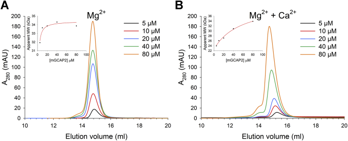
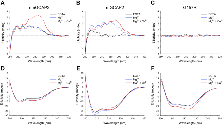
In murine and bovine photoreceptors, guanylate cyclase-activating protein 2 (GCAP2) activates retinal guanylate cyclases (GCs) at low Ca2+ levels, thus contributing to the Ca2+/cGMP negative feedback on the cyclase together with its paralog guanylate cyclase-activating protein 1, which has the same function but different Ca2+ sensitivity. In humans, a GCAP2 missense mutation (G157R) has been associated with inherited retinal degeneration (IRD) via an unknown molecular mechanism. Here, we characterized the biochemical properties of human GCAP2 and the G157R variant, focusing on its dimerization and the Ca2+/Mg2+-binding processes in the presence or absence of N-terminal myristoylation. We found that human GCAP2 and its bovine/murine orthologs significantly differ in terms of oligomeric properties, cation binding, and GC regulation. Myristoylated GCAP2 endothermically binds up to 3 Mg2+ with high affinity and forms a compact dimer that may reversibly dissociate in the presence of Ca2+. Conversely, nonmyristoylated GCAP2 does not bind Mg2+ over the physiological range and remains as a monomer in the absence of Ca2+. Both myristoylated and nonmyristoylated GCAP2 bind Ca2+ with high affinity. At odds with guanylate cyclase-activating protein 1 and independently of myristoylation, human GCAP2 does not significantly activate retinal GC1 in a Ca2+-dependent fashion. The IRD-associated G157R variant is characterized by a partly misfolded, molten globule-like conformation with reduced affinity for cations and prone to form aggregates, likely mediated by hydrophobic interactions. Our findings suggest that GCAP2 might be mostly implicated in processes other than phototransduction in human photoreceptors and suggest a possible molecular mechanism for G157R-associated IRD.

Keywords: GCAP; GUCA1B; cGMP; calcium-binding proteins; guanylate cyclase (guanylyl cyclase); neurodegenerative disease; phototransduction; retina; retinal degeneration; vision.


Conflict of interest statement

Conflict of interest The authors declare that they have no conflicts of interest with the contents of this article.

Figures

Figure 1
Figure 1
Three-dimensional structure of nmGCAP2 and assessment of the molecular mass of GCAP2 variants by MALDI-TOF MS.A, Cartoon representation of the homology model of human nmGCAP2 (built on bovine nmGCAP2 template, PDB entry 1JBA (8)), colored in a red-to-purple rainbow according to the sequence. Ca2+ are displayed as red spheres, and IRD-associated R157 variant is shown as white sticks with N atoms highlighted in blue. EF1 to EF4 are labeled, together with residue R157. The protein view is rotated by 90° clockwise along the x-axis in the right panel. MALDI spectra of (B) nmGCAP2, (C) mGCAP2, and (D) G157R with the molecular mass corresponding to each peak. GCAP2, guanylate cyclase–activating protein 2; IRD, inherited retinal dystrophy; mGCAP2, myristoylated GCAP2; nmGCAP2, nonmyristoylated GCAP2.
Figure 2
Figure 2
Effects of Mg2+and Ca2+on the apparent molecular mass and the hydrodynamic diameter of GCAP2 variants. Analytical SEC profiles of ∼40-μM (A) nmGCAP2 and (B) mGCAP2 in the presence of 500-μM EGTA + 1-mM Mg2+ (blue) or 1-mM Mg2+ + 500-μM Ca2+ (red). Hydrodynamic diameter estimation by DLS of ∼40-μM (C) nmGCAP2 and (D) mGCAP2 in the presence of 500-μM EGTA + 1-mM Mg2+ (blue) or 1-mM Mg2+ + 500-μM Ca2+ (red). DLS, dynamic light scattering; GCAP2, guanylate cyclase–activating protein; mGCAP2, myristoylated GCAP2; nmGCAP2, nonmyristoylated GCAP2; SEC, size-exclusion chromatography.
Figure 3
Figure 3
Effects of Mg2+andCa2+on mGCAP2 dimerization. Analytical gel-filtration chromatograms of 5-μM (black), 10-μM (red), 20-μM (blue), 40-μM (green), and 80-μM (orange) mGCAP2 in the presence of (A) 500-μM EGTA + 1-mM Mg2+ and (B) 1-mM Mg2+ + 500-μM Ca2+. Insets show the apparent molecular mass as a function of the mGCAP2 concentration together with the optimal fitting to a hyperbolic curve (see Experimental procedures). GCAP2, guanylate cyclase–activating protein 2; mGCAP2, myristoylated GCAP2.
Figure 4
Figure 4
Conformational changes of GCAP2 variants upon cation binding monitored by CD spectroscopy. Near-UV CD spectra of 30-μM (A) nmGCAP2, (B) mGCAP2, and (C) G157R in the presence of 500-μM EGTA (black) and after addition of 1-mM Mg2+ (blue) and 1-mM Ca2+ (red). Far-UV CD spectra of 8-μM (D) nmGCAP2, (E) mGCAP2, and (F) G157R in the presence of 300-μM EGTA (black) and after addition of 1-mM Mg2+ (blue) and 600-μM Ca2+ (red). GCAP2, guanylate cyclase–activating protein 2; mGCAP2, myristoylated GCAP2; nmGCAP2, nonmyristoylated GCAP2.
Figure 5
Figure 5
Cation-dependent thermal stability of GCAP2 variants monitored by CD spectroscopy. Thermal denaturation profiles of 8-μM (A) nmGCAP2, (B) mGCAP2, and (C) G157R in the presence of 300-μM EGTA (black), 300-μM EGTA + 1-mM Mg2+ (blue) and 1-mM Mg2+ + 600-μM Ca2+ (red). When possible, profiles were fitted to a 4-parameter Hill sigmoid, yielding the Tm values reported in Table 2. GCAP2, guanylate cyclase–activating protein 2; mGCAP2, myristoylated GCAP2; nmGCAP2, nonmyristoylated GCAP2.
Figure 6
Figure 6
Conformational changes of GCAP2 variants upon ion binding assessed by proteolytic cleavage. Limited proteolysis of GCAP2 variants after 10-min incubation with trypsin (60:1 molar ratio) in the presence of 1-mM EDTA, 500-μM EGTA + 1-mM Mg2+, or 1-mM Mg2+ + 1-mM Ca2+. GCAP2, guanylate cyclase–activating protein 2; mGCAP2, myristoylated GCAP2; nmGCAP2, nonmyristoylated GCAP2.
Figure 7
Figure 7
Variations in hydrophobicity of GCAP2 variants upon cation binding investigated by ANS fluorescence. Fluorescence spectra of 30-μM ANS and 2-μM (A) nmGCAP2, (B) mGCAP2, and (C) G157R in the presence of 500-μM EGTA (black) and after sequential additions of 1-mM Mg2+ (blue) and 1-mM Ca2+ (red). The spectrum of ANS is shown as a green dashed line in panel A. ANS, anilinonaphthalene-1-sulfonic acid; GCAP2, guanylate cyclase–activating protein 2; mGCAP2, myristoylated GCAP2; nmGCAP2, nonmyristoylated GCAP2.
Figure 8
Figure 8
Effects of Mg2+and Ca2+on the electrophoretic mobility of GCAP2 variants. SDS-PAGE of 20-μM GCAP2 variants in the presence of 5-mM EDTA, 5-mM EGTA + 1-mM free Mg2+, or 1-mM Mg2+ + 1-mM Ca2+. GCAP2, guanylate cyclase–activating protein 2; mGCAP2, myristoylated GCAP2; nmGCAP2, nonmyristoylated GCAP2.
Figure 9
Figure 9
Metal cations binding to GCAP2 variants measured by ITC. Representative ITC titrations of 20-μM nmGCAP2 (A–C) and mGCAP2 (D–F). Mg2+ titrations of (A) nmGCAP2 and (D) mGCAP2. Ca2+ titrations of (B) nmGCAP2 and (E) mGCAP2 in the absence and in the presence (C) and (F) of 1-mM Mg2+. Upper panels show heat pulses, and lower panels represent molar enthalpy changes relative to each injection. When possible, data were fitted to a two- or three-sequential binding sites (see Experimental procedures), yielding apparent dissociation constants (KD), enthalpy changes (ΔH), and entropy changes (−TΔS) reported in Tables 3 and 4. GCAP2, guanylate cyclase–activating protein 2; ITC, isothermal titration calorimetry; mGCAP2, myristoylated GCAP2; nmGCAP2, nonmyristoylated GCAP2.

Similar articles

Cited by

References

    1. Koch K.W., Dell'Orco D. Protein and signaling networks in vertebrate photoreceptor cells. Front. Mol. Neurosci. 2015;8:67. - PMC - PubMed
    1. Palczewski K., Polans A.S., Baehr W., Ames J.B. Ca(2+)-binding proteins in the retina: Structure, function, and the etiology of human visual diseases. Bioessays. 2000;22:337–350. - PubMed
    1. Koch K.W., Stryer L. Highly cooperative feedback control of retinal rod guanylate cyclase by calcium ions. Nature. 1988;334:64–66. - PubMed
    1. Hwang J.Y., Koch K.W. Calcium- and myristoyl-dependent properties of guanylate cyclase-activating protein-1 and protein-2. Biochemistry. 2002;41:13021–13028. - PubMed
    1. Peshenko I.V., Moiseyev G.P., Olshevskaya E.V., Dizhoor A.M. Factors that determine Ca2+ sensitivity of photoreceptor guanylyl cyclase. Kinetic analysis of the interaction between the Ca2+-bound and the Ca2+-free guanylyl cyclase activating proteins (GCAPs) and recombinant photoreceptor guanylyl cyclase 1 (RetGC-1) Biochemistry. 2004;43:13796–13804. - PubMed

Publication types

MeSH terms