

Lymphoma in Psittacine Birds: A Histological and Immunohistochemical Assessment
- PMID: 33813951
- PMCID: PMC8290990
- DOI: 10.1177/03009858211002180
Lymphoma in Psittacine Birds: A Histological and Immunohistochemical Assessment
Abstract
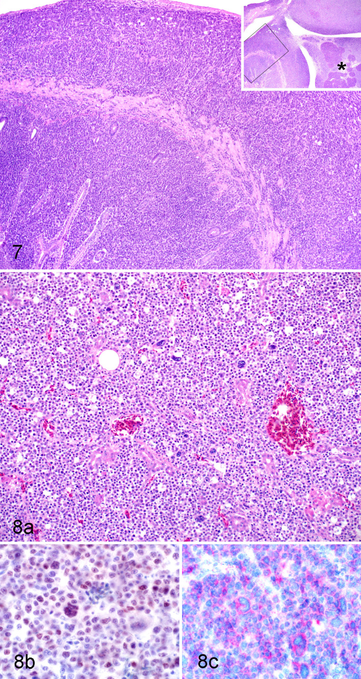
In psittacine birds, round cell neoplasms that originate from lymphocytes, plasma cells, histiocytes, or mast cells are sporadic and poorly described. The lack of morphological and immunohistochemical diagnostic criteria or grading schemes make specific diagnoses and prognoses challenging. We assessed cases of psittacine birds diagnosed with round cell neoplasia from 3 North American veterinary diagnostic laboratories to describe the diagnostic features of these tumors. For all cases, demographic data, anatomic distribution, histological features, and immunoreactivity for T (CD3) and B (Pax5 and MUM-1) cell markers were assessed using tissue microarrays and whole slide mounts. Thirty-eight psittacine birds representing 14 species were included. Tumors were mainly infiltrative and multicentric, were composed of homogenous sheets of round to polygonal cells, and commonly presented with a high mitotic count (average 21 mitoses per high-power field). Based on Pax5 immunoreactivity, B-cell lymphoma was most common (19/38 [50%]), and was significantly associated with involvement of the gastrointestinal and urogenital systems. Of the 38 cases, 6 (16%) were consistent with T-cell lymphoma, 3 (8%) with plasma cell tumor, and 3 (8%) were double-reactive for both B- and T-lymphocyte markers. This is the first study to describe morphologic and immunohistochemical features of round cell neoplasia in a large number of psittacine birds, and provides benchmark data for future studies aimed at elucidating the diagnosis and prognosis of these neoplasms. These data also provide useful information about reactivity of commercially available antibodies as lymphocyte markers in tissues of multiple psittacine species.
Keywords: Psittaciformes; avian; histopathology; immunohistochemistry; immunophenotype; lymphoma; round cell neoplasia.
Conflict of interest statement
Figures


References
-
- Aquino SM, Hamor RE, Valli VE, et al. Progression of an orbital T-cell rich B-cell lymphoma to a B-cell lymphoma in a dog. Vet Pathol. 2000;37(5):465–469. - PubMed
-
- Burgos-Rodríguez AG, Garner M, Ritzman TK, et al. Cutaneous lymphosarcoma in a double yellow-headed Amazon parrot (Amazona ochrocephala oratrix). J Avian Med Surg. 2007;21(4):283–289. - PubMed
-
- Calnek BW. Pathogenesis of Marek’s disease virus infection. In: Hirai K, ed. Marek’s Disease. Springer-Verlag; 2001:25–55. - PubMed
-
- Carrasco V, Rodríguez-Bertos A, Rodríguez-Franco F, et al. Distinguishing intestinal lymphoma from inflammatory bowel disease in canine duodenal endoscopic biopsy samples. Vet Pathol. 2015;52(4):668–675. - PubMed
-
- Coleman C. Lymphoid neoplasia in pet birds: a review. J Avian Med Surg. 1995;9(1):3–7.
Publication types
MeSH terms
LinkOut - more resources
Full Text Sources
Other Literature Sources
Medical
