Review
. 2021 Apr 5;15(1):181.
doi: 10.1186/s13256-021-02761-1.

Bilateral pelvic kidneys with upper pole fusion and malrotation: a case report and review of the literature

Affiliations
Review

Bilateral pelvic kidneys with upper pole fusion and malrotation: a case report and review of the literature

Hussam S Khougali et al. J Med Case Rep. .

Abstract

Background: The incidence of ectopic kidneys is 1:12,000 clinically and 1:900 postmortem. Patients with pelvic mal-rotated kidneys are more susceptible to recurrent urinary tract infections, recurrent renal stones, and renal injury. Fusion of the kidney lower poles is relatively common compared to other types of renal anomalies.

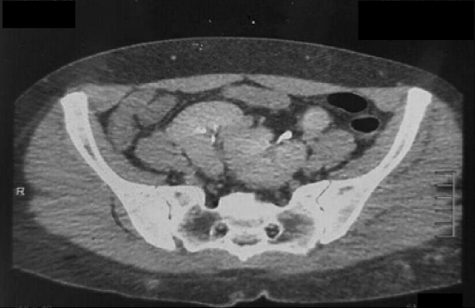
Case presentation: We present the case of a 36-year-old Sudanese female patient who presented with a long history of recurrent urinary tract infections unresponsive to antibiotics. Ultrasound scan revealed bilateral pelvic kidneys. Computed tomography (CT) urography confirmed bilateral ectopic fused kidneys, with the left kidney mal-rotated (renal pelvis facing upwards and laterally). Kidney infection secondary to vesicoureteral reflux was diagnosed. Antibiotics were prescribed according to culture and sensitivity. The patient responded well to ciprofloxacin.

Conclusion: A history of recurrent urinary tract infections without an apparent cause is highly suggestive of renal anomaly and should be investigated expediently. Ultrasonography or CT imaging may be utilized to aid in diagnosis. Early recognition may help prevent the high risk of end-stage renal failure associated with anomalies.

Keywords: Bilateral pelvic kidneys; Renal ectopia; Renal reversed rotation.


Conflict of interest statement

The authors declare that they have no competing interests.

Figures

Fig. 1
Fig. 1
Abdominopelvic ultrasound scan showed ectopic kidneys at the hemi-pelvis, fused in their upper poles, normal size and texture of the kidneys with normal corticomedullary differentiation, no stones or obstructive changes
Fig. 2
Fig. 2
Computed tomography urography. The right kidney is ectopically placed in the pelvis, measures 9.6 cm bipolar length, and is medially and inferiorly faced. The left kidney is also ectopically placed in the pelvis, measures 9.3 cm in bipolar length, is mal-rotated as the pelvis faces upward and laterally, both kidneys are partially fused at their upper poles
Fig. 3
Fig. 3
Computed tomography urography. The right kidney is ectopically placed in the pelvis, measures 9.6 cm bipolar length, and is medially and inferiorly faced. The left kidney is also ectopically placed in the pelvis, measures 9.3 cm in bipolar length, and is mal-rotated as the pelvis faces upward and laterally, both kidneys are partially fused at their upper poles
Fig. 4
Fig. 4
Computed tomography urography. The right kidney is ectopically placed in the pelvis, measures 9.6 cm bipolar length, and is medially and inferiorly faced. The left kidney is also ectopically placed in the pelvis, measures 9.3 cm in bipolar length, and is mal-rotated as the pelvis faces upward and laterally, both kidneys are partially fused at their upper poles

References

    1. Meizner I, Yitzhak M, Levi A, Barki Y, Barnhard Y, Glezerman M. Fetal pelvic kidney: a challenge in prenatal diagnosis? Ultrasound Obstet Gynecol. 1995;5:391–393. doi: 10.1046/j.1469-0705.1995.05060391.x. - DOI - PubMed
    1. Rodriguez MM. Congenital anomalies of the kidney and the urinary tract (CAKUT) Fetal Pediatr Pathol. 2014;33:293–320. doi: 10.3109/15513815.2014.959678. - DOI - PMC - PubMed
    1. Türkvatan A, Olçer T, Cumhur T. Multidetector CT urography of renal fusion anomalies. Diagn Interv Radiol. 2009;15:127–134. - PubMed
    1. Pohl HG, Belman AB. Congenital anomalies of the urinary tract. Curr Pediatr Rev. 2014 doi: 10.2174/157339631002140513102152. - DOI - PubMed
    1. Iseko KI, Iseko JBR, Ewuzie OC, Akpan CI. Bilateral, fused pelvic, ectopic, laterally rotated kidneys: a case report. Radiol Case Rep. 2020;15(7):841–845. doi: 10.1016/j.radcr.2020.04.012. - DOI - PMC - PubMed

LinkOut - more resources