. 2021 Jan;31(Suppl 1):S70-S79.
doi: 10.4103/ijri.IJRI_967_20. Epub 2021 Jan 23.

Role of chest radiography in the management of COVID-19 pneumonia: An overview and correlation with pathophysiologic changes

Affiliations

Role of chest radiography in the management of COVID-19 pneumonia: An overview and correlation with pathophysiologic changes

Anirudh Kohli et al. Indian J Radiol Imaging. 2021 Jan.

Abstract

Background: Chest radiography (CXR) is a widely available baseline radiological modality in evaluating symptomatic patients with suspected or confirmed Covid-19 disease. Serial changes can help in monitoring the patients in conjunction with the clinical status of these patients in a hospital setting.

Purpose: The purpose of this study was to analyse the patterns of radiological findings on chest radiograph (CXR) for suspected and confirmed COVID-19 patients on initial presentation to the emergency medical services (EMS) on admission and to assess the progression and resolution.

Materials and methods: In this study, patients who presented to EMS of a multispeciality hospital as suspected or confirmed Covid-19 on consecutive reverse transcriptase polymerase chain reaction (RT-PCR). CXR was examined for findings of haziness, patterns and distribution of opacities. Progression and regression of findings in serial CXR were studied and evaluated with the clinical and laboratory parameters. High resolution CT (HRCT) chest was performed initially for some patients.

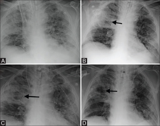
Results: 756 RT-PCR confirmed COVID-19 patients were included in our study who had initial CXR. 510 (67.46%) of our patients with positive initial RT-PCR showed abnormal baseline CXR. The abnormal findings were described as haziness akin to ground glass opacities (GGO) on CT, peripheral opacities, patchy parenchymal opacities and consolidation. Peripheral opacities and lower zone distribution were the commonest pattern of CXR abnormalities with bilateral involvement. The severity of findings on serial CXR and radiographic regression was studied along with follow-up to assess response to treatment. Forty-six patients showed features of acute lung injury (ALI). Complications and new CXR findings were reported for patients who were given ventilator support.

Conclusion: CXR is a valuable baseline radiological investigation on hospital admission in symptomatic patients with suspected or confirmed Covid-19 presenting to the EMS as it helps to monitor the progress and regression of the disease in conjunction with clinical findings.

Keywords: Acute lung injury; Covid-19 pneumonia; ventilator-associated pneumonia.


Conflict of interest statement

There are no conflicts of interest.

Figures

Figure 1
Figure 1
Chest radiograph PA view of RT-PCR proven COVID positive patient showing patchy opacities in bilateral mid and lower zones predominantly involving the peripheral lung fields (left more than right)
Figure 2
Figure 2
Chest radiograph PA view of RT-PCR proven COVID positive patient shows showing ill-defined air space consolidations with reticular opacities (black arrows) in bilateral mid and lower zones
Figure 3
Figure 3
Chest radiograph of a COVID positive patient showing patchy nodular lesions in bilateral mid and lower zones representing atypical appearance
Figure 4
Figure 4
Chest radiograph of COVID positive patient proven by RT-PCR showing subtle hazy opacities (black arrow) in right lower zone
Figure 5
Figure 5
Chest radiograph of COVID positive patient proven by RT-PCR shows dense airspace consolidation in subpleural region of right upper and lower zones
Figure 6
Figure 6
Chest radiograph of COVID positive patient proven on RT-PCR shows small nodular consolidations involving both lung fields representing broncho-pneumonia pattern
Figure 7
Figure 7
Chest radiograph in a Covid positive patient proven on RT-PCR reveals diffuse airspace consolidation in bilateral mid and lower zones (right more than left lung)
Figure 8 (A and B)
Figure 8 (A and B)
(A) Chest radiograph of a RT-PCR proven COVID positive patient shows no abnormality. (B) HRCT chest was done on same day as patient was symptomatic revealed patchy areas of ground glass densities with interlobular septal thickening in posterior basal segment of right lower lobe and medial basal segment of left lower lobe
Figure 9 (A-D)
Figure 9 (A-D)
Serial chest radiographs over 7 days in a patient with Covid-19 infection depicting (A-C) progression of the disease in the first 3 days with (D) gradual resolution of opacities over the next 4 days
Figure 10
Figure 10
Serial chest radiographs over 4 days in a patient with Covid-19 infection showing progression of the disease for which patient was intubated. Follow-up radiograph demonstrates resolution on the 4th day
Figure 11 (A and B)
Figure 11 (A and B)
Serial chest radiographs at 7 days interval in a patient with Covid-19 infection showing resolution of the diffuse consolidation involving the bilateral mid and lower zones in Chest X-ray (A)
Figure 12 (A-D)
Figure 12 (A-D)
Serial chest radiographs in RT-PCR proven COVID positive patient showing progression of the ill-defined hazy opacities noted in right lower zone (A) with increase in densities of opacities and involvement of bilateral mid lower zones (B and C) and gradual regression in the densities of these opacities noted in last chest X-ray (D)
Figure 13
Figure 13
Serial chest radiographs in a patient on mechanical ventilation. Diffuse airspace consolidation involving right lung field and left mid and lower zone. There is resolution of the opacities visualised in right upper and bilateral mid zones
Figure 14
Figure 14
Serial chest radiographs over 5 days in a case with COVID pneumonia showing progression of density and area of airspace opacities. Patient was intubated on the 5th day and unfortunately expired one day later
Figure 15
Figure 15
Portable chest radiograph of COVID pneumonia patient with diffuse airspace opacities in bilateral lung fields with relative sparing of left upper zone. Patient was intubated and put on positive ventilation because of diffuse lung involvement. Linear lucencies in right mid zone (red arrow) representing pulmonary interstitial emphysema
Figure 16 (A-F)
Figure 16 (A-F)
Serial chest radiographs (A-F) in a COVID positive patient who presented with acute breathlessness. (A) Normal initial OPD Chest radiograph. (B-F) Subsequent Chest radiographs show progression of the patchy ground glass opacities to diffuse consolidation for which patient required ventilatory support. (F) Mediastinal emphysema (black arrow) is noted as a result of barotrauma
Figure 17
Figure 17
Portable chest X-ray of a COVID pneumonia patient with bilateral diffuse involvement of lungs developed right-sided pneumothorax with collapse of underlying right lung and mediastinal shift towards left due to barotrauma. Incidentally noted is a well-defined oval radiolucency representing a pneumatocele in right lower zone (black arrow)
Figure 18
Figure 18
Portable Chest radiograph after chest tube insertion in Covid positive patient with bilateral diffuse air space consolidation requiring positive pressure ventilation developed left sided pneumothorax likely due to barotrauma resulting in collapse of the underlying left lung and mediastinal shift towards right. A well-defined oval radioluceny in periphery of left mid zone representing a pneumatocele (black arrow) with linear radiolucencies in left mid and lower zones around heart representing pulmonary interstitial emphysema
Figure 19
Figure 19
Portable chest X-ray in a Covid pneumonia patient on mechanical ventilation developed mediastinal emphysema (black arrow) and diffuse subcutaneous emphysema as a result of barotrauma. Linear radiolucencies noted in right mid zone representing pulmonary interstitial emphysema. Incidentally noted is central line coiled back in left IJV (red arrow)
Figure 20 (A-D)
Figure 20 (A-D)
Serial portable chest radiographs in a Covid positive patient requiring mechanical ventilation, showing (A) dense consolidation in right upper and mid zone (this was a new finding as compared to old X-rays). (B-D) shows cavitation in dense consolidation. Endotracheal tube swab (ETS) grew Acinetobacter Baumanii on culture. Pneumonia resolved on appropriate antibiotic therapy as shown in follow up chest X-rays
Figure 21 (A-D)
Figure 21 (A-D)
Serial portable chest radiographs in a Covid positive patient. (A) showing airspace consolidation in right lung and left mid and lower zone (B) resolution in the density and extent of airspace consolidation in right lung field and left mid zone (C) ill-defined consolidation in right mid and lower zone (new finding-red arrow). Microbiological investigations revealed Acinetobacter Baumanii on culture (D) shows resolution of extent and density of opacities noted in right mid and lower zones after appropriate antibiotic treatment

References

    1. Jacobi A, Chung M, Bernheim A, Eber C. Portable chest X-ray in coronavirus disease-19 (COVID-19): A pictorial review. Clin Imaging. 2020;64:35–42. - PMC - PubMed
    1. Cevik M, Kuppalli K, Kindrachuk J, Peiris M. Virology, transmission, and pathogenesis of SARS-CoV-2? BMJ. 2020;371:m3862. doi: 10.1136/bmj.m3862. - PubMed
    1. Rodrigues JC, Hare SS, Edey A, Devaraj A, Jacob J, Johnstone A, et al. An update on COVID-1 9 for the radiologist-A British society of Thoracic Imaging statement. Clin Radiol. 2020;75:323–5. - PMC - PubMed
    1. Tang X, Du R, Wang R, Cao TZ, Guan LL, Yang CQ, et al. Comparison of hospitalized patients with ARDS caused by covid-19 and H1N1. Chest. 2020;158(1):195–205. - PMC - PubMed
    1. Wong HY, Lam HY, Fong AH, Leung ST, Chin TW, Lo CS, et al. Frequency and distribution of chest radiographic findings in COVID-19 positive patients. Radiology. 2020;296(No. 2):201160. - PMC - PubMed