

Pattern recognition of high-resolution computer tomography (HRCT) chest to guide clinical management in patients with mild to moderate COVID-19
- PMID: 33814769
- PMCID: PMC7996691
- DOI: 10.4103/ijri.IJRI_774_20
Pattern recognition of high-resolution computer tomography (HRCT) chest to guide clinical management in patients with mild to moderate COVID-19
Abstract
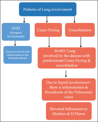
Aim: To describe the distribution of lung patterns determined by High Resolution Computed Tomography (HRCT) in COVID patients with mild and moderate lung involvement and outcomes after early identification and management with steroids and anticoagulants.
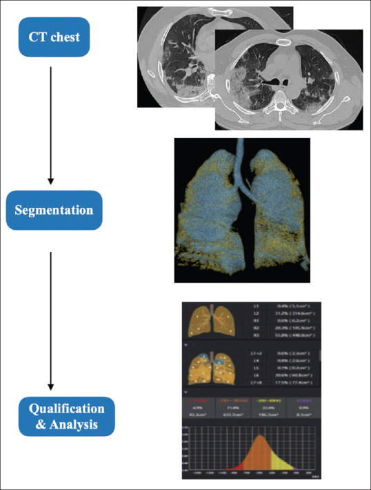
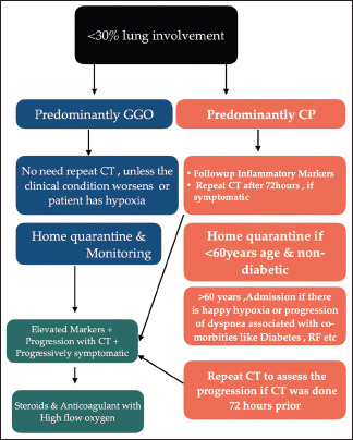
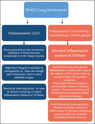
Material and methods: A cross sectional study of COVID-19 patients with mild and moderate lung involvement presenting at 5 healthcare centres in Trichy district of South TamilNadu in India. Patients underwent HRCT to assess patterns and severity of lung involvement, Inflammatory markers (LDH/Ferritin) and D-Dimer assay and clinical correlation with signs and symptoms. Patients were assessed for oxygen, steroid and anticoagulant therapy, clinical recovery or progression on follow up and details on mortality were collected. The RSNA, Fleischer Society guidelines and CORADS score was used for radiological reporting. New potential classification of patterns of percentage of lung parenchyma involvement in Covid patients is being suggested.
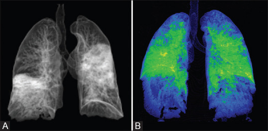
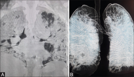
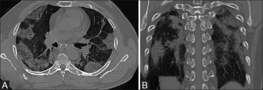
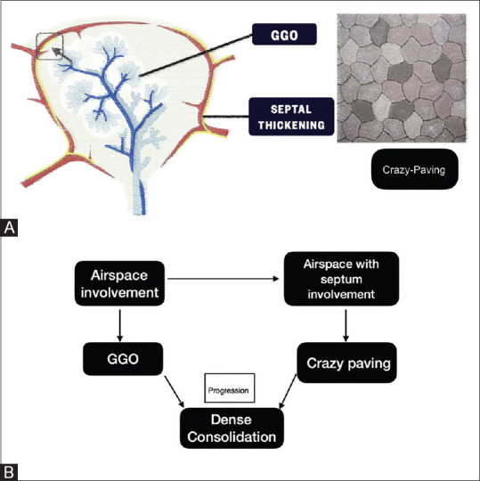
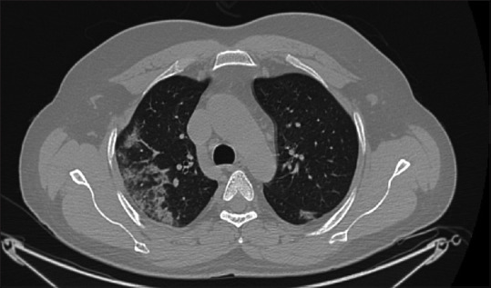
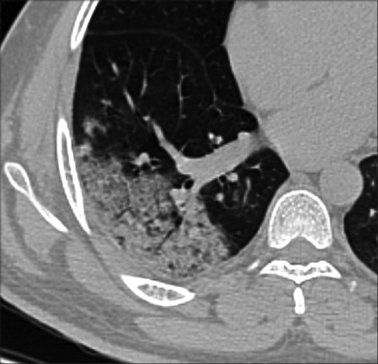
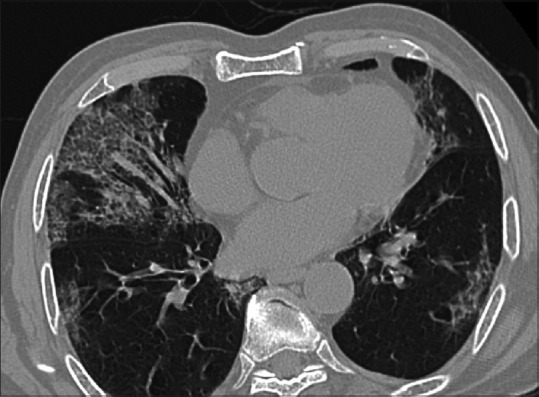
Results: The study included 7,340 patients with suspected COVID and 3,963 (53.9%) patients had lung involvement based on HRCT. RT PCR was positive in 74.1% of the CT Positive cases. Crazy Pavement pattern was predominant (n = 2022, 51.0%) and Ground Glass Opacity (GGO) was found in 1,941 (49.0%) patients in the study. Severe lung involvement was more common in the Crazy Pavement pattern. Patients with GGO in moderate lung involvement were significantly more likely to recover faster compared to Crazy Pavement pattern (P value <0.001).
Conclusion: HRCT chest and assessment of lung patterns can help triage patients to home quarantine and hospital admission. Early initiation of steroids and anticoagulants based on lung patterns can prevent progression to more severe stages and aid early recovery. HRCT can play a major role to triage and guide management especially as RT PCR testing and results are delayed for the benefit of patients and in a social cause to decrease the spread of the virus.
Keywords: COVID 19; HRCT; crazy pavement pattern; ground glass opacity.
Copyright: © 2021 Indian Journal of Radiology and Imaging.
Conflict of interest statement
There are no conflicts of interest.
Figures
Similar articles
-
Pattern of Lung Involvement in Predicting Severity and Sequelae in Patients With COVID-19.Cureus. 2022 Dec 26;14(12):e32973. doi: 10.7759/cureus.32973. eCollection 2022 Dec. Cureus. 2022. PMID: 36712734 Free PMC article.
-
[Spatial and temporal distribution and predictive value of chest CT scoring in patients with COVID-19].Zhonghua Jie He He Hu Xi Za Zhi. 2021 Mar 12;44(3):230-236. doi: 10.3760/cma.j.cn112147-20200522-00626. Zhonghua Jie He He Hu Xi Za Zhi. 2021. PMID: 33721937 Chinese.
-
An Analysis of High-Resolution Computed Tomography Chest Manifestations of COVID-19 Patients in Pakistan.Cureus. 2020 Jul 24;12(7):e9373. doi: 10.7759/cureus.9373. Cureus. 2020. PMID: 32850241 Free PMC article.
-
Predictors of the chest CT score in COVID-19 patients: a cross-sectional study.Virol J. 2021 Nov 18;18(1):225. doi: 10.1186/s12985-021-01699-6. Virol J. 2021. PMID: 34794467 Free PMC article. Review.
-
Similarities and Differences of Early Pulmonary CT Features of Pneumonia Caused by SARS-CoV-2, SARS-CoV and MERS-CoV: Comparison Based on a Systemic Review.Chin Med Sci J. 2020 Sep 30;35(3):254-261. doi: 10.24920/003727. Chin Med Sci J. 2020. PMID: 32972503 Free PMC article.
Cited by
-
Pattern of Lung Involvement in Predicting Severity and Sequelae in Patients With COVID-19.Cureus. 2022 Dec 26;14(12):e32973. doi: 10.7759/cureus.32973. eCollection 2022 Dec. Cureus. 2022. PMID: 36712734 Free PMC article.
-
Use of serum KL-6 and chest radiographic severity grade to predict 28-day mortality in COVID-19 patients with pneumonia: a retrospective cohort study.BMC Pulm Med. 2024 Apr 18;24(1):187. doi: 10.1186/s12890-024-02992-0. BMC Pulm Med. 2024. PMID: 38637771 Free PMC article.
References
-
- Hansell DM, Bankier AA, MacMahon H, McLoud TC, Müller NL, Remy J. Fleischner society: Glossary of terms for thoracic imaging. Radiology. 2008;246:697–722. 10.1148/radiol.2462070712. - PubMed
-
- Salehi S, Abedi A, Balakrishnan S, Gholamrezanezhad A. Coronavirus disease 2019 (COVID-19): A systematic review of imaging findings in 919 patients. AJR Am J Roentgenol. 2020:1–7. 10.2214/AJR.20.23034. - PubMed
-
- Prokop M, van Everdingen W, van Rees Vellinga T, Quarles van Ufford J, Stöger L, Beenen L, Geurts B, Gietema H, Krdzalic J, Schaefer-Prokop C, van Ginneken B, Brink M. CO-RADS – A categorical CT-assessment scheme for patients with suspected COVID-19: Definition and evaluation. Radiology. 2020 doi: 10.1148/radiol.2020201473. - PMC - PubMed
LinkOut - more resources
Full Text Sources
Other Literature Sources