

Role of Cervical Cancer Biomarkers p16 and Ki67 in Abnormal Cervical Cytological Smear
- PMID: 33814802
- PMCID: PMC7960869
- DOI: 10.1007/s13224-020-01380-y
Role of Cervical Cancer Biomarkers p16 and Ki67 in Abnormal Cervical Cytological Smear
Abstract
Introduction: Cervical cancer is the most common cancer in India. Screening for cervical cancer helps in marked reduction of invasive cervical cancers. The low sensitivity of Papanicolaou cytology (Pap smear) and high-risk human papillomavirus (HR-HPV) in excluding high-grade intraepithelial lesion (ASC-H) leads to unnecessary referrals to colposcopy-guided biopsy. The combined cervical cytology screening and HR-HPV have its own limitations and still need further standardization. Using additional biomarkers like staining with p16 and Ki-67 might help in triaging abnormal pap smear.
Materials and methods: A prospective, cross-sectional study was performed over a period of 16 months in the Department of Obstetrics and Gynaecology, in collaboration with Department of Pathology. Study was conducted to know the efficacy of immunostaining with p16/Ki-67 in predicting the presence of significant lesion in cases of mild cytological atypia. PAP smears (conventional and LBC) along with P16, Ki-67 and available biopsies were correlated.
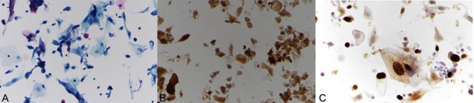
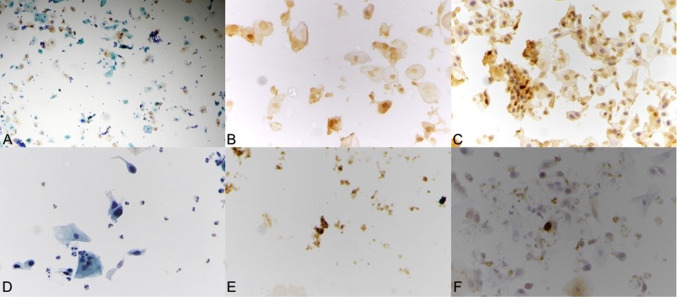
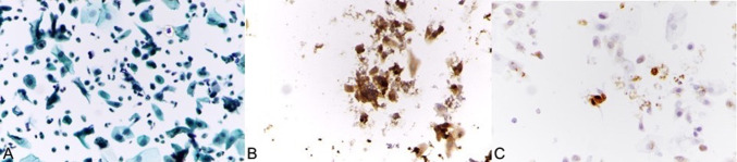
Results: Liquid-based cytology (LBC) was done in 2134 cases, out of which 46 cases showed abnormal cytological findings such as [22 atypical squamous cells of undetermined significance (ASCUS), 3 low-grade squamous intraepithelial lesion (LSIL), 8 atypical squamous cells cannot exclude high-grade lesion (ASC-H), 6 high-grade squamous intraepithelial lesion (HSIL), 5 squamous cell carcinoma (SCC), 2 adenocarcinoma, 1 atypical glandular cells of undetermined significance (AGUS)]. Immunostaining with p16 and Ki-67 was performed on 38 cases of abnormal cytological smears. Out of 38 abnormal cytology cases, 28 cases had shown co-staining for both p16 and Ki-67, suggestive of true HPV infection of the cells. Of the 38 cases, 07/14 ASCUS, 06/06 HSIL, 07/08 ASC-H, 05/05 squamous cell carcinoma and 02/02 adenocarcinoma also showed dual positivity for p16 and Ki-67. One case of AGUS was diagnosed, but the smear was unsatisfactory for immunocytochemical evaluation and excluded from the study. Three cases of LSIL were also diagnosed on cytological evaluation, and 1 of them however showed positivity for p16 and Ki-67 on immunocytochemistry (ICC). In the ASC-US group, the sensitivity and specificity of the immunostaining in diagnosing CIN2 + lesions were 87.51%, and in LSIL group, the sensitivity and specificity of the immunostaining in diagnosing CIN2 + lesions were 100%. p16/Ki-67 positivity also increased with cytological severity which in turn corresponded with histological findings: it reached from 50% in ASC-US to 100% in both HSIL and SCC categories.
Conclusion: This immunostaining with p16 and Ki67 can be a useful method in the triaging of the ASC-US and the LSIL group as considering the high sensitivity and specificity values.
Keywords: ASC-US; ICC; Ki67; Liquid-based cytology; p16.
© Federation of Obstetric & Gynecological Societies of India 2020.
Conflict of interest statement
Conflict of interestNone declared.
Figures
References
LinkOut - more resources
Full Text Sources
Research Materials
Miscellaneous