

Nodular Posterior Scleritis Masquerading as a Subretinal Mass
- PMID: 33814821
- PMCID: PMC7993044
- DOI: 10.4103/meajo.MEAJO_216_19
Nodular Posterior Scleritis Masquerading as a Subretinal Mass
Abstract
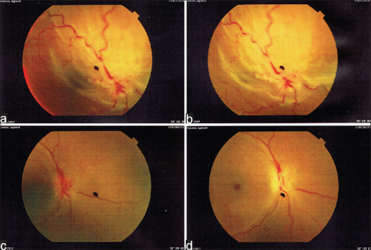
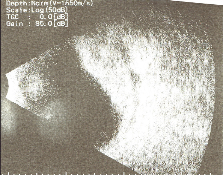
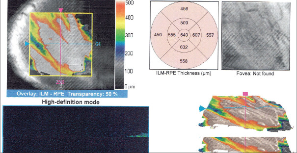
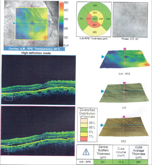
This is the case report of a 50-year-old female with no systemic comorbidities who presented to the eye clinic with a 1-month history of right-sided eye pain and visual loss. Examination revealed no signs of inflammation in the right eye, with no proptosis or conjunctival injection. A relative afferent pupillary defect was present with no inflammatory cells in the vitreous. On fundoscopy, there was a swollen disc, a large superior creamy white subretinal mass associated with a shallow overlying retinal detachment. B-scan ultrasonography confirmed the presence of a subretinal mass. Hematological investigations revealed an elevated erythrocyte sedimentation rate. Infective and autoimmune markers were negative. A diagnosis was made of nodular posterior scleritis and the patient was treated with intravenous corticosteroids initially, and subsequently switched to oral corticosteroids. There was complete resolution of the mass with optic atrophy as a result. Posterior nodular scleritis is an extremely rare potentially vision-threatening ocular condition that requires multimodal investigations to diagnose and treat appropriately.
Keywords: Nodular scleritis; posterior scleritis; subretinal mass.
Copyright: © 2021 Middle East African Journal of Ophthalmology.
Conflict of interest statement
There are no conflicts of interest.
Figures
Similar articles
-
Multimodal Imaging of Nodular Posterior Scleritis: Case Report and Review of the Literature.Middle East Afr J Ophthalmol. 2020 Jul 20;27(2):134-138. doi: 10.4103/meajo.MEAJO_115_20. eCollection 2020 Apr-Jun. Middle East Afr J Ophthalmol. 2020. PMID: 32874049 Free PMC article. Review.
-
Posterior scleritis in children: clinical features and treatment.Ophthalmology. 2012 Jan;119(1):59-65. doi: 10.1016/j.ophtha.2011.09.030. Epub 2011 Dec 3. Ophthalmology. 2012. PMID: 22137553
-
NODULAR POSTERIOR SCLERITIS: Clinico-Sonographic Characteristics and Proposed Diagnostic Criteria.Retina. 2016 Feb;36(2):392-401. doi: 10.1097/IAE.0000000000000699. Retina. 2016. PMID: 26296144
-
Nodular Posterior Scleritis Mimicking Choroidal Tumor in a Patient With Systemic Lupus Erythematous: A Case Report and Literature Review.Asia Pac J Ophthalmol (Phila). 2016 Sep-Oct;5(5):324-9. doi: 10.1097/APO.0000000000000165. Asia Pac J Ophthalmol (Phila). 2016. PMID: 26692258 Review.
-
Giant nodular posterior scleritis simulating choroidal melanoma.Indian J Ophthalmol. 2006 Jun;54(2):120-2. doi: 10.4103/0301-4738.25835. Indian J Ophthalmol. 2006. PMID: 16770031
References
-
- McCluskey PJ, Watson PG, Lightman S, Haybittle J, Restori M, Branley M. Posterior scleritis: Clinical features, systemic associations, and outcome in a large series of patients. Ophthalmology. 1999;106:2380–6. - PubMed
-
- Agrawal R, Lavric A, Restori M, Pavesio C, Sagoo MS. Nodular posterior scleritis: Clinico-sonographic characteristics and proposed diagnostic criteria. Retina. 2016;36:392–401. - PubMed
-
- Tanaka R, Kaburaki T, Ohtomo K, Takamoto M, Komae K, Numaga J, et al. Clinical characteristics and ocular complications of patients with scleritis in Japanese. Jpn J Ophthalmol. 2018;62:517–24. - PubMed
-
- Shukla D, Kim R. Giant nodular posterior scleritis simulating choroidal melanoma. Indian J Ophthalmol. 2006;54:120–2. - PubMed
-
- Sainz de la Maza M, Jabbur NS, Foster CS. Severity of scleritis and episcleritis. Ophthalmology. 1994;101:389–96. - PubMed
Publication types
MeSH terms
Substances
LinkOut - more resources
Full Text Sources
