. 2021 Mar 19:14:647054.
doi: 10.3389/fnmol.2021.647054. eCollection 2021.

Tau: A Signaling Hub Protein

Affiliations

Tau: A Signaling Hub Protein

Rebecca L Mueller et al. Front Mol Neurosci. .

Abstract

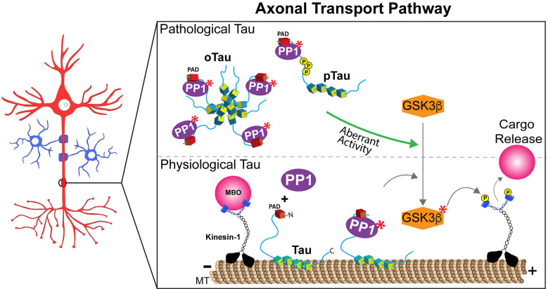
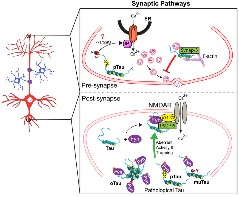
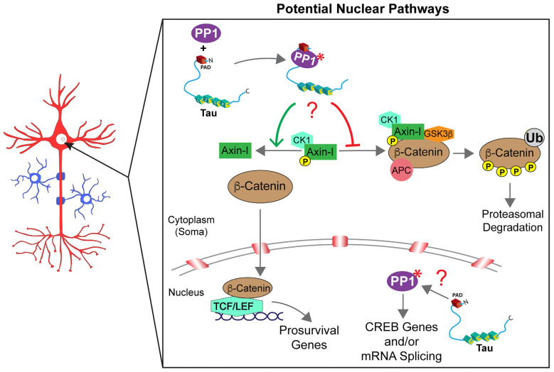
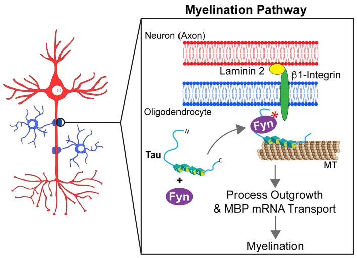
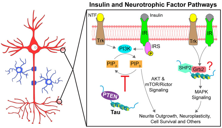
Over four decades ago, in vitro experiments showed that tau protein interacts with and stabilizes microtubules in a phosphorylation-dependent manner. This observation fueled the widespread hypotheses that these properties extend to living neurons and that reduced stability of microtubules represents a major disease-driving event induced by pathological forms of tau in Alzheimer's disease and other tauopathies. Accordingly, most research efforts to date have addressed this protein as a substrate, focusing on evaluating how specific mutations, phosphorylation, and other post-translational modifications impact its microtubule-binding and stabilizing properties. In contrast, fewer efforts were made to illuminate potential mechanisms linking physiological and disease-related forms of tau to the normal and pathological regulation of kinases and phosphatases. Here, we discuss published work indicating that, through interactions with various kinases and phosphatases, tau may normally act as a scaffolding protein to regulate phosphorylation-based signaling pathways. Expanding on this concept, we also review experimental evidence linking disease-related tau species to the misregulation of these pathways. Collectively, the available evidence supports the participation of tau in multiple cellular processes sustaining neuronal and glial function through various mechanisms involving the scaffolding and regulation of selected kinases and phosphatases at discrete subcellular compartments. The notion that the repertoire of tau functions includes a role as a signaling hub should widen our interpretation of experimental results and increase our understanding of tau biology in normal and disease conditions.

Keywords: axon; kinase; nucleus; oligodendrocyte; phosphatase; scaffold protein; synpase; tauopathy.


Conflict of interest statement

The authors declare that the research was conducted in the absence of any commercial or financial relationships that could be construed as a potential conflict of interest.

Figures

Figure 1
Figure 1
Tau regulates anterograde fast axonal transport through a PP1-GSK3β pathway. Lower panel: Exposure of a phosphatase activating domain (PAD, red domain) located at the N-terminus of tau causes activation of protein phosphatase 1 (PP1, in purple). Active PP1 (labeled by red asterisks) dephosphorylates and activates glycogen synthase kinase 3β (GSK3β). Active GSK3β (labeled by red asterisks) phosphorylates light chain subunits of kinesin-1, an event that promotes the release of membrane-bound organelle (MBO) cargoes transported by this major motor protein. Spatially discrete post-translational modifications in tau that either promote (i.e., phosphorylation events in the proline-rich region) or prevent (i.e., N-terminal phosphorylation) PAD exposure may thus allow the localized delivery of selected MBOs at functionally distinct axonal subdomains, such as en passant pre-synaptic terminals or nodes of Ranvier under normal physiological conditions. Although depicted bound to microtubules (MT), tau’s dynamic interaction with MTs in normal conditions likely provides opportunities for tau to interact with PP1 (and/or other enzymes) on or off the MT surface. Upper panel: By extension, aberrant PAD exposure associated pathological forms of tau such as aggregates/oligomers (oTau), mutant tau, and selected pathological post-translational modifications (e.g., phosphorylation, pTau), may disrupt homeostatic maintenance of this process.
Figure 2
Figure 2
Tau regulates several signaling synaptic pathways. Upper panel: At the pre-synaptic compartment, tau undergoes phosphorylation at the AT8 epitope (pTau). This event promotes PAD exposure, which in turn increases Ca2+ release from intracellular stores and reduces neurotransmitter release in an IP3-dependent manner, which might involve PP1 and/or GSK3 (indicated by a red question mark). Also, pathological forms of tau reportedly restrict vesicle mobility through scaffold interactions with the vesicular protein synaptogyrin-3 and F-actin. Lower panel: Tau localizes Fyn to the post-synapse allowing Fyn to phosphorylate NMDAR2b at Y1472, which leads to NMDAR stabilization via interaction with PSD-95. Pathological forms of tau including aggregated/oligomeric tau (oTau), specific phosphorylated forms of tau (pTau), or mutant tau (muTau) can affect tau-Fyn binding, ultimately leading to aberrant synaptic activity by promoting increased trapping of Fyn in dendritic spines.
Figure 3
Figure 3
Tau may control multiple signaling pathways in the nucleus. β-catenin degradation/signaling is regulated by the assembly or disassembly of a cytoplasmic destruction complex [composed of glycogen synthase kinase-3β (GSK3β), casein kinase 1 (CK1), adenomatous polyposis coli (APC), and axin-I]. Whether β-catenin is free to translocate into the nucleus (anti-apoptotic conditions) or degraded in the cytoplasm through ubiquitination (Ub; in pro-apoptotic conditions) is driven, at least in part, by the phosphorylation status of axin-I. PP1 directly regulates axin-I phosphorylation with PP1-dependent dephosphorylation of axin-I preventing the formation of the destruction complex and favoring anti-apoptotic β-catenin signaling in the nucleus. Through interactions via its PAD domain, cytoplasmic tau may activate PP1 (marked by a red asterisk) and promote axin-I dephosphorylation (indicated by a red question mark), but this potential mechanism awaits further investigation. Similarly, the co-location of tau and PP1 within the nucleus suggests a potential role may exist for tau to modulate intra-nuclear PP1-dependent pathways such as cAMP response element-binding protein phosphorylation and signaling and/or mRNA splicing. However, future studies are needed to fully elucidate potential intra-nuclear tau signaling mechanisms (indicated by a red question mark).
Figure 4
Figure 4
Tau modulates Fyn kinase pathways in myelinating oligodendrocytes. Myelination requires the outgrowth of oligodendrocyte processes and the formation of contact points between oligodendrocytes and axons. For this to occur, the mRNA of the myelin basic protein (MBP) needs to be targeted to those contact points. Tau acts as a scaffold protein linking Fyn kinase to the microtubule (MT) cytoskeleton of oligodendrocyte processes. Upon establishment of contact points at the neuron-glia interface, a signaling cascade involving tau and Fyn kinase is activated. That signaling pathway culminates into the rearrangement of the cell cytoskeleton that leads MBP mRNA to myelination sites and promotes the growth of oligodendrocyte processes.
Figure 5
Figure 5
Tau promotes signal transduction of insulin and neurotrophic factor pathways. Neurotrophic factors (NTF) and insulin activate specific receptor tyrosine kinases (Trks and IRs, respectively). Following autophosphorylation, the receptors initiate signaling through multiple downstream pathways, two of which are partially modulated by tau. Activation of AKT signaling by PI3K is regulated by PIP3 levels, increased through phosphorylation by PI3K and decreased through dephosphorylation by PTEN. Tau reportedly binds to PTEN directly to inhibit its phosphatase activity (red inhibitory arrow), thereby promoting AKT activation. Tau can also promote MAPK signaling through an interaction with SHP2, a tyrosine phosphatase downstream of the receptor tyrosine kinases. Tau also interacts with the SH3 domain of Grb2, an activator of SHP2 but whether tau, SHP2, and Grb2 form a complex and the downstream effects are not known (indicated by a red question mark). Phosphorylation at specific residues may impact the modulatory role of tau on these pathways.

References

    1. Ahmed T., Van der Jeugd A., Blum D., Galas M. C., D’Hooge R., Buee L., et al. . (2014). Cognition and hippocampal synaptic plasticity in mice with a homozygous tau deletion. Neurobiol. Aging 35, 2474–2478. 10.1016/j.neurobiolaging.2014.05.005 - DOI - PubMed
    1. Belkadi A., LoPresti P. (2008). Truncated Tau with the Fyn-binding domain and without the microtubule-binding domain hinders the myelinating capacity of an oligodendrocyte cell line. J. Neurochem. 107, 351–360. 10.1111/j.1471-4159.2008.05600.x - DOI - PubMed
    1. Benetatos J., Bennett R. E., Evans H. T., Ellis S. A., Hyman B. T., Bodea L. G., et al. . (2020). PTEN activation contributes to neuronal and synaptic engulfment by microglia in tauopathy. Acta Neuropathol. 140, 7–24. 10.1007/s00401-020-02151-9 - DOI - PMC - PubMed
    1. Bhaskar K., Yen S.-H., Lee G. (2005). Disease-related modifications in tau affect the interaction between Fyn and Tau. J. Biol. Chem. 280, 35119–35125. 10.1074/jbc.M505895200 - DOI - PubMed
    1. Black M. M. (2016). Axonal transport: the orderly motion of axonal structures. Methods Cell Biol. 131, 1–19. 10.1016/bs.mcb.2015.06.001 - DOI - PubMed

LinkOut - more resources