. 2021 Mar 18:12:622084.
doi: 10.3389/fphys.2021.622084. eCollection 2021.

Absence of Rgs5 Influences the Spatial and Temporal Fluctuation of Cardiac Repolarization in Mice

Affiliations

Absence of Rgs5 Influences the Spatial and Temporal Fluctuation of Cardiac Repolarization in Mice

Zi-Liang Song et al. Front Physiol. .

Abstract

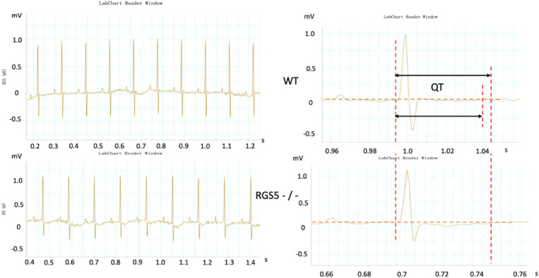
Aims: This study investigated the contribution of the regulator of G-protein signaling 5 (Rgs5) knockout to the alteration of the action potential duration (APD) restitution and repolarizing dispersion in ventricle.

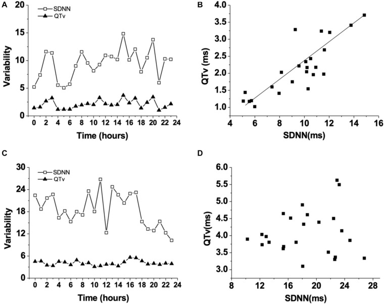
Methods and results: The effects of Rgs5-/- were investigated by QT variance (QTv) and heart rate variability analysis of Rgs5-/- mice. Monophasic action potential analysis was investigated in isolated Rgs5-/- heart. Rgs5-/- did not promote ventricular remodeling. The 24-h QTv and QT variability index (QTVI) of the Rgs5-/- mice were higher than those of wild-type (WT) mice (P < 0.01). In WT mice, a positive correlation was found between QTv and the standard deviation of all NN intervals (r = 0.62; P < 0.01), but not in Rgs5-/- mice (R = 0.01; P > 0.05). The absence of Rgs5 resulted in a significant prolongation of effective refractory period and APD in isolated ventricle. In addition, compared with WT mice, the knockout of Rgs5 significantly deepened the slope of the APD recovery curve at all 10 sites of the heart (P < 0.01) and increased the spatial dispersions of Smax (COV-Smax) (WT: 0.28 ± 0.03, Rgs5-/-: 0.53 ± 0.08, P < 0.01). Compared with WT heart, Rgs5-/- increased the induced S1-S2 interval at all sites of heart and widened the window of vulnerability of ventricular tachyarrhythmia (P < 0.05).

Conclusion: Our findings indicate that Rgs5-/- is an important regulator of ventricular tachyarrhythmia in mice by prolonging ventricular repolarization and increasing spatial dispersion in ventricle.

Keywords: G-protein signaling 5; QT variability; cardiac repolarization; spatial dispersion; ventricular arrhythmic.


Conflict of interest statement

The authors declare that the research was conducted in the absence of any commercial or financial relationships that could be construed as a potential conflict of interest.

Figures

FIGURE 1
FIGURE 1
Fibrosis assessment of Rgs5–/– and WT ventricles. The mRNA expression levels of fibrotic mediators including Tgfb1, Col1a1, and Col3a1 were analyzed in the ventricles of WT and Rgs5–/– mice (A). PSR staining was performed on the histological sections of the ventricles in WT and Rgs5–/– mice (B).
FIGURE 2
FIGURE 2
Representative electrocardiograms were recorded in WT and RGS5–/– mice. Results of QT intervals measurement were displayed by waveform.
FIGURE 3
FIGURE 3
Changes of QT variability (QTv) and SDNN in WT mice during 24 h (A). Correlation between QT variability (QTv) and SDNN in WT mice (B). Changes of QT variability (QTv) and SDNN in Rgs5–/– mice during 24 h (C). Correlation between QT variability (QTv) and SDNN in Rgs5–/– mice (D).
FIGURE 4
FIGURE 4
Representative APD was recorded in WT and RGS5–/– mice.
FIGURE 5
FIGURE 5
Dispersion of ventricular repolarization in isolated mouse ventricle. Action potential duration (APD) (A) and effective refractory period (ERP) (C) at different sites of ventricle in WT and Rgs5–/– mice. COV-APD90 (B) and COV-ERP (D) at different sites in WT and Rgs5–/– mice. Left ventricular anterior base (LAB), left ventricle anterior middle wall (LAM), left ventricle posterior base (LPB),left atrial appendage (LAA), left ventricle posterior middle wall(LPM), left ventricular posterior apex(LPA), right ventricular outflow tract(RVOT),right ventricular base(RB), right ventricular middle wall (RM), and right ventricular apex (RA).
FIGURE 6
FIGURE 6
Representative examples of APD restitution curves at each site in WT and Rgs5–/– mice.
FIGURE 7
FIGURE 7
Smax (A) at different sites of ventricle in WT and Rgs5–/– mice. COV-Smax (B) at different sites in WT and Rgs5–/– mice.
FIGURE 8
FIGURE 8
S1–S2 interval-induced ventricular tachyarrhythmia at different sites of ventricle in WT and Rgs5–/– mice (A). Window of vulnerability (WOV) of VA in 10 sites of ventricle in Rgs5–/– and WT hearts (B).

References

    1. Antzelevitch C. (2007). Role of spatial dispersion of repolarization in inherited and acquired sudden cardiac death syndromes. Am. J. Physiol. Heart Circ. Physiol. 293 H2024–H2038. 10.1152/ajpheart.00355.2007 - DOI - PMC - PubMed
    1. Arai K., Nakagawa Y., Iwata T., Horiguchi H., Murata K. (2013). Relationships between QT interval and heart rate variability at rest and the covariates in healthy young adults. Auton. Neurosci. 173 53–57. 10.1016/j.autneu.2012.11.006 - DOI - PubMed
    1. Badran H. M., Elnoamany M. F., Soltan G., Ezat M., Elsedi M., Abdelfatah R. A., et al. (2012). Relationship of mechanical dyssynchrony to QT interval prolongation in hypertrophic cardiomyopathy. Eur. Heart J. Cardiovasc. Imaging 13 423–432. 10.1093/ejechocard/jer290 - DOI - PubMed
    1. Coronel R., Wilms-Schopman F. J., Opthof T., Janse M. J. (2009). Dispersion of repolarization and arrhythmogenesis. Heart Rhythm 6 537–543. 10.1016/j.hrthm.2009.01.013 - DOI - PubMed
    1. Decker K. F., Heijman J., Silva J. R., Hund T. J., Rudy Y. (2009). Properties and ionic mechanisms of action potential adaptation, restitution, and accommodation in canine epicardium. American journal of physiology. Am. J. Physiol. Heart Circ. Physiol. 296 H1017–H1026. 10.1152/ajpheart.01216.2008 - DOI - PMC - PubMed

LinkOut - more resources