. 2021 Mar 17:12:599149.
doi: 10.3389/fendo.2021.599149. eCollection 2021.

Androgen and Luteinizing Hormone Stimulate the Function of Rat Immature Leydig Cells Through Different Transcription Signals

Affiliations

Androgen and Luteinizing Hormone Stimulate the Function of Rat Immature Leydig Cells Through Different Transcription Signals

Xiaoheng Li et al. Front Endocrinol (Lausanne). .

Abstract

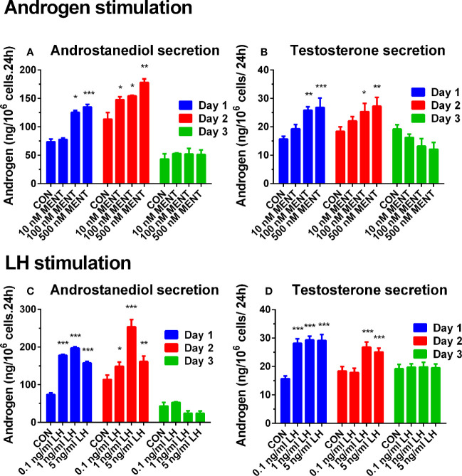
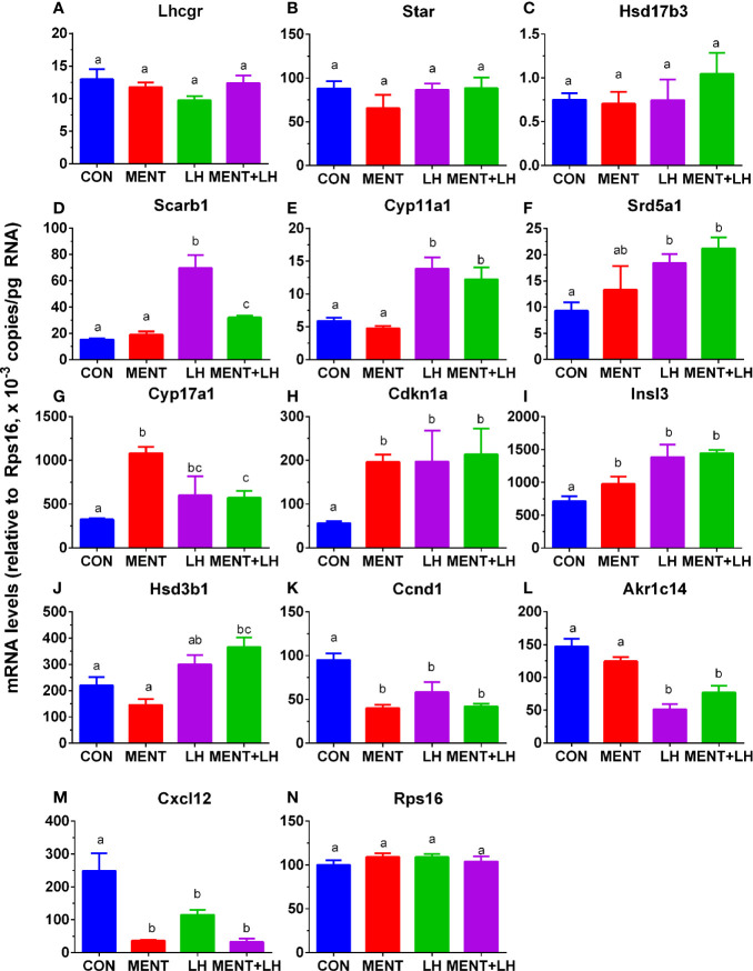
The function of immature Leydig cells is regulated by hormones, such as androgen and luteinizing hormone (LH). However, the regulation of this process is still unclear. The objective of this study was to determine whether luteinizing hormone (LH) or androgens contribute to this process. Immature Leydig cells were purified from 35-day-old male Sprague Dawley rats and cultured with LH (1 ng/ml) or androgen (7α-methyl-19- nortestosterone, MENT, 100 nM) for 2 days. LH or MENT treatment significantly increased the androgens produced by immature Leydig cells in rats. Microarray and qPCR and enzymatic tests showed that LH up-regulated the expression of Scarb1, Cyp11a1, Cyp17a1, and Srd5a1 while down-regulated the expression of Sult2a1 and Akr1c14. On the contrary, the expression of Cyp17a1 was up-regulated by MENT. LH and MENT regulate Leydig cell function through different sets of transcription factors. We conclude that LH and androgens participate in the regulation of rat immature Leydig cell function through different transcriptional pathways.

Keywords: development; immature Leydig cell; luteinizing hormone; regulation; steroidogenesis; testosterone.


Conflict of interest statement

The authors declare that the research was conducted in the absence of any commercial or financial relationships that could be construed as a potential conflict of interest.

Figures

Figure 1
Figure 1
Effect of hormone treatment on androgen production in rat immature Leydig cells. Immature rat Leydig cells were cultured with MENT or LH for 3 days. The levels of medium testosterone and androstanediol were measured. (A, B) MENT treatment. (C, D) LH treatment. (A, C) Androstanediol level. (B, D) Testosterone level. Mean ± SEM, n = 6. *, **, and *** represent significant differences compared with the control group each day at P < 0.05, 0.01, and 0.001, respectively.
Figure 2
Figure 2
Regimen of treatment and effect of MENT and LH alone or in combination on androgen production in immature Leydig cells in rats. Immature rat Leydig cells were cultured with MENT or LH alone or in combination for 2 days or with FLU or MENT or in combination for another study (A). The levels of medium androstanediol and testosterone were measured. (A) Regimen for this experiment and for experiment in Figures 7 and 8 . (B) Androstanediol. (C) Testosterone level. Mean ± SEM, n = 4. The same letter means that there is no significant difference between the two groups at P < 0.05.
Figure 3
Figure 3
Changes in genes overlap between LH and MENT. (A) 98 genes up-regulated by MENT ≥3 times, 74 genes up-regulated by LH ≥3 times, 1 gene down-regulated by LH and 14 unaltered genes; (B) 67 genes down-regulated by MENT were 18 genes down-regulated, 4 genes up-regulated and 44 unaltered genes after LH treatment.
Figure 4
Figure 4
Analysis of gene pathways for MENT, LH alone or in combination (MENT+LH) to regulate steroidogenesis. Red indicates up-regulated genes; green indicates down-regulated genes. Solid arrow indicates androgen synthesis pathway; Dash arrow indicates steroid metabolism; Double arrows indicates transport of cholesterol.
Figure 5
Figure 5
Transcription factors are regulated by MENT, LH alone or in combination (MENT+LH). Red indicates that the gene is up-regulated; green indicates that the gene is down-regulated. (A) Up-regulated expression of genes. (B) Down-regulated expression of genes; Four categories: (1) Only up-regulated/down-regulated in MENT-treated groups; (2) Up-regulated/down-regulated in MENT alone or MENT+LH groups; (3) Only up-regulated/down-regulated in LH-treated group; (4) Up-regulated/down-regulated in MENT, LH, and MENT + LH groups.
Figure 6
Figure 6
Messenger RNA levels are regulated by MENT or LH alone or in combination (MENT+LH) after qPCR analysis. In the presence of MENT or LH alone or in combination, immature Leydig cells were cultured for 2 days. The following genes were analyzed (A–N): Lhcgr, Star, Hsd17b3, Scarb1, Cyp11a1, Srd5a1, Cyp17a1, Cdkn1a, Insl3, Hsd3b1, Ccnd1, Akr1c14, Cxcl12, and Rps16. The data are shown as mean ± SEM, n = 4–8 preparations. The same letter indicates that the difference between the two groups is not statistically significant (P < 0.05).
Figure 7
Figure 7
Antagonistic effect of flutamide (FLU) on MENT-induced gene up-regulation. In the presence of MENT, FLU alone or in combination (MENT +FLU), immature Leydig cels were cultured for 2 days. The following genes were analyzed (A–D): Cyp17a1, Cdkn1a, Svs5, and Ptgds. The data are shown as mean ± SEM, n = 4–8 preparations. The same letter indicates that the difference between the two groups is not statistically significant (P < 0.05).
Figure 8
Figure 8
Androgen receptor antagonist flutamide (FLU) antagonizes MENT-induced CYP17A1 level. In the presence of MENT, FLU alone or in combination (MENT +FLU), immature Leydig cells were cultured for 2 days. Analysis of CYP17A1 and actin β (ACTB) levels. The CYP17A1 level was adjusted to ACTB. (A) Western blot; (B) Quantitative data. The data is shown as mean ± SEM, n = 3 preparations. The same letter indicates that the difference between the two groups is not statistically significant (P < 0.05).
Figure 9
Figure 9
Effect of MENT, LH alone or in combination (MENT+LH) on the activity of steroidogenic enzymes in immature Leydig cells. In the presence of MENT, LH alone or in combination (MENT+LH), immature Leydig cells were cultured for 2 days. (A–F): CYP11A1, HSD3B1, CYP17A1, HSD17B3, SRD5A1, AKR1C14, respectively. The data is shown as mean ± SEM, n = 4 preparations. The same letter indicates that the difference between the two groups is not statistically significant (P < 0.05).

References

    1. Chen P, Zirkin BR, Chen H. Stem Leydig Cells in the Adult Testis: Characterization, Regulation and Potential Applications. Endocr Rev (2020) 41:22–32. 10.1210/endrev/bnz013 - DOI - PMC - PubMed
    1. Haider SG. Cell biology of Leydig cells in the testis. Int Rev Cytol (2004) 233:181–241. 10.1016/S0074-7696(04)33005-6 - DOI - PubMed
    1. Ye L, Li X, Li L, Chen H, Ge RS. Insights into the development of the adult Leydig cell lineage from stem Leydig cells. Front Physiol (2017) 8:430. 10.3389/fphys.2017.00430 - DOI - PMC - PubMed
    1. Ge RS, Dong Q, Sottas CM, Papadopoulos V, Zirkin BR, Hardy MP. In search of rat stem Leydig cells: identification, isolation, and lineage-specific development. Proc Natl Acad Sci U.S.A. (2006) 103:2719–24. 10.1073/pnas.0507692103 - DOI - PMC - PubMed
    1. Stanley E, Lin CY, Jin S, Liu J, Sottas CM, Ge R, et al. . Identification, proliferation, and differentiation of adult Leydig stem cells. Endocrinology (2012) 153:5002–10. 10.1210/en.2012-1417 - DOI - PMC - PubMed

Publication types

MeSH terms

LinkOut - more resources