Review
. 2021 Mar 18:12:662017.
doi: 10.3389/fendo.2021.662017. eCollection 2021.

Astrocyte Clocks and Glucose Homeostasis

Affiliations
Review

Astrocyte Clocks and Glucose Homeostasis

Olga Barca-Mayo et al. Front Endocrinol (Lausanne). .

Abstract

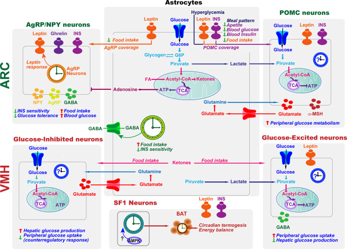
The endogenous timekeeping system evolved to anticipate the time of the day through the 24 hours cycle of the Earth's rotation. In mammals, the circadian clock governs rhythmic physiological and behavioral processes, including the daily oscillation in glucose metabolism, food intake, energy expenditure, and whole-body insulin sensitivity. The results from a series of studies have demonstrated that environmental or genetic alterations of the circadian cycle in humans and rodents are strongly associated with metabolic diseases such as obesity and type 2 diabetes. Emerging evidence suggests that astrocyte clocks have a crucial role in regulating molecular, physiological, and behavioral circadian rhythms such as glucose metabolism and insulin sensitivity. Given the concurrent high prevalence of type 2 diabetes and circadian disruption, understanding the mechanisms underlying glucose homeostasis regulation by the circadian clock and its dysregulation may improve glycemic control. In this review, we summarize the current knowledge on the tight interconnection between the timekeeping system, glucose homeostasis, and insulin sensitivity. We focus specifically on the involvement of astrocyte clocks, at the organism, cellular, and molecular levels, in the regulation of glucose metabolism.

Keywords: astrocytes; circadian clock; diabetes; glucose homeostasis; metabolism.


Conflict of interest statement

The authors declare that the research was conducted in the absence of any commercial or financial relationships that could be construed as a potential conflict of interest.

Figures

Figure 1
Figure 1
The circadian timing system. The timekeeping system is composed of two pacemakers (the SCN and the FEO), and peripheral clocks in other brain areas and peripheral tissues. Light inputs reaching the SCN via the retina and the retinohypothalamic tract, are the most important Zeitgeber for the SCN, which in turn synchronizes peripheral clocks through neural, endocrine, temperature, and behavioral signals. Feeding-related signals (INS and ketones bodies) generated by peripheral tissues and IGLNPY neurons entrain the FEO, which regulate the outputs such as FAA.
Figure 2
Figure 2
Core molecular clock network. The mammalian molecular clock consists of a transcriptional-translational feedback loop involving the clock proteins CLOCK, ARNTL, PER, and CRY and the nuclear receptors REV-ERB and ROR. The positive limb (CLOCK and BMAL1) heterodimerizes and activates the transcription of downstream genes, including Per, Cry, Rorα and Rev-erb. The negative limb proteins (PERs and CRYs) multimerize and inhibit CLOCK/BMAL1. In a secondary loop, Bmal1 and Clock are regulated by the repressor REV-ERB and its opposing nuclear receptor RORα, which bind competitively to the shared element RORE, thus repressing or activating the transcription of the Bmal1 or Clock gene, respectively. Post-translational modifications of core-clock factors can also regulate transcription (e.g., deacetylation of BMAL1 or PER2 by SIRT1, BMAL1 phosphorylation or PER2 translation by INS signaling; O- GlcNAcylation of CLOCK, BMAL1 and PER2; and CRY1 phosphorylation by AMPK).
Figure 3
Figure 3
Astrocytes and clocks modulate hypothalamic glucose-sensing mechanisms. The ability of POMC and NPY/AgRP neuronal populations in ARC to alter energy metabolism is due to their sensitivity to several circulating signals, including hormones, such as leptin and insulin (INS), and nutrients. Hypothalamic astrocytes provide neurons with structural support and nutrients. Moreover, hyperglycemia, INS, and leptin signaling in this glial cell type lead to changes in the astrocytic coverage of POMC and/or AgRP neurons to regulate glucose sensing. Glucose transported into astrocytes can be metabolized to lactate, which is released and taken up by neurons and metabolized into pyruvate to serve as a glycolytic substrate. Astrocytes can also modulate synaptic transmission by uptake of neurotransmitters from the synaptic cleft (glutamate and GABA) and by releasing gliotransmitters such as adenosine, which inhibits AgRP neurons. In the ARC, the astrocyte circadian clock might control food intake and glucose homeostasis by regulating the uptake of GABA. On the other hand, the clock in AgRP neurons is required for coordinating leptin response and glucose metabolism. VMH neurons include glucose-sensing cells, referred to as glucose-excited and glucose-inhibited neurons. The activation of glucose-excited neurons leads to decreased hepatic glucose production and increased peripheral glucose uptake. VMH glucose-inhibited neurons are activated in response to hypoglycemia. In recurrent hypoglycemia, high accumulation of lactate enhances the glucose-excited neuronal activity and consequent GABA release, inhibiting the counterregulatory response. A high-fat diet increases astrocyte ketone bodies production, which are exported to VMH neurons to ultimately control food intake. Subsets of VMH neurons also express SF1. These SF1 neurons contain a clock that modulates energy expenditure by regulating cyclic thermogenesis in brown adipose tissue (BAT). As hypothalamic AMPK modulates BAT thermogenesis (116), and has a crucial role in the molecular clock, it would be interesting to investigate its involvement in the control of rhythmic BAT thermogenesis by the SF1 neuronal clock. TCA, tricarboxylic acid cycle.

References

    1. Hastings MH, Maywood ES, Reddy AB. Two decades of circadian time. J Neuroendocrinol (2008) 20:812–9. 10.1111/j.1365-2826.2008.01715.x - DOI - PubMed
    1. Moore RY, Eichler VB. Loss of a circadian adrenal corticosterone rhythm following suprachiasmatic lesions in the rat. Brain Res (1972) 42:201–6. 10.1016/0006-8993(72)90054-6 - DOI - PubMed
    1. Stephan FK, Zucker I. Circadian rhythms in drinking behavior and locomotor activity of rats are eliminated by hypothalamic lesions. Proc Natl Acad Sci U S A (1972) 69:1583–6. 10.1073/pnas.69.6.1583 - DOI - PMC - PubMed
    1. Nagoshi E, Saini C, Bauer C, Laroche T, Naef F, Schibler U. Circadian Gene Expression in Individual FibroblastsCell-Autonomous and Self-Sustained Oscillators Pass Time to Daughter Cells. Cell (2004) 119:693–705. 10.1016/s0092-8674(04)01054-2 - DOI - PubMed
    1. Welsh DK, Yoo SH, Liu AC, Takahashi JS, Kay SA. Bioluminescence imaging of individual fibroblasts reveals persistent, independently phased circadian rhythms of clock gene expression. Curr Biol (2004) 14:2289–95. 10.1016/j.cub.2004.11.057 - DOI - PMC - PubMed

Publication types

LinkOut - more resources