A Therapeutic Strategy to Combat HIV-1 Latently Infected Cells With a Combination of Latency-Reversing Agents Containing DAG-Lactone PKC Activators
- PMID: 33815322
- PMCID: PMC8010149
- DOI: 10.3389/fmicb.2021.636276
A Therapeutic Strategy to Combat HIV-1 Latently Infected Cells With a Combination of Latency-Reversing Agents Containing DAG-Lactone PKC Activators
Abstract
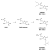
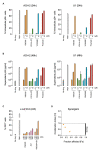
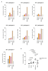
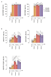
Advances in antiviral therapy have dramatically improved the therapeutic effects on HIV type 1 (HIV-1) infection. However, even with potent combined antiretroviral therapy, HIV-1 latently infected cells cannot be fully eradicated. Latency-reversing agents (LRAs) are considered a potential tool for eliminating such cells; however, recent in vitro and in vivo studies have raised serious concerns regarding the efficacy and safety of the "shock and kill" strategy using LRAs. In the present study, we examined the activity and safety of a panel of protein kinase C (PKC) activators with a diacylglycerol (DAG)-lactone structure that mimics DAG, an endogenous ligand for PKC isozymes. YSE028, a DAG-lactone derivative, reversed HIV-1 latency in vitro when tested using HIV-1 latently infected cells (e.g., ACH2 and J-Lat cells) and primary cells from HIV-1-infected individuals. The activity of YSE028 in reversing HIV-1 latency was synergistically enhanced when combined with JQ1, a bromodomain and extra-terminal inhibitor LRA. DAG-lactone PKC activators also induced caspase-mediated apoptosis, specifically in HIV-1 latently infected cells. In addition, these DAG-lactone PKC activators showed minimal toxicity in vitro and in vivo. These data suggest that DAG-lactone PKC activators may serve as potential candidates for combination therapy against HIV-1 latently infected cells, especially when combined with other LRAs with a different mechanism, to minimize side effects and achieve maximum efficacy in various reservoir cells of the whole body.
Keywords: HIV-1; HIV-1 latently infected cells; HIV-1 reservoirs; diacylglycerol-lactone; protein kinase C activator.
Copyright © 2021 Matsuda, Kobayakawa, Kariya, Tsuchiya, Ryu, Tsuji, Ishii, Gatanaga, Yoshimura, Okada, Hamada, Mitsuya, Tamamura and Maeda.
Conflict of interest statement
The authors declare that the research was conducted in the absence of any commercial or financial relationships that could be construed as a potential conflict of interest.
Figures

Similar articles
-
Synthesis and evaluation of DAG-lactone derivatives with HIV-1 latency reversing activity.Eur J Med Chem. 2023 Aug 5;256:115449. doi: 10.1016/j.ejmech.2023.115449. Epub 2023 May 3. Eur J Med Chem. 2023. PMID: 37224601 Free PMC article.
-
Benzolactam-related compounds promote apoptosis of HIV-infected human cells via protein kinase C-induced HIV latency reversal.J Biol Chem. 2019 Jan 4;294(1):116-129. doi: 10.1074/jbc.RA118.005798. Epub 2018 Nov 9. J Biol Chem. 2019. PMID: 30413535 Free PMC article.
-
Discovery of Potent DAG-Lactone Derivatives as HIV Latency Reversing Agents.ACS Infect Dis. 2024 Jun 14;10(6):2250-2261. doi: 10.1021/acsinfecdis.4c00194. Epub 2024 May 21. ACS Infect Dis. 2024. PMID: 38771724
-
Current Status of Latency Reversing Agents Facing the Heterogeneity of HIV-1 Cellular and Tissue Reservoirs.Front Microbiol. 2020 Jan 24;10:3060. doi: 10.3389/fmicb.2019.03060. eCollection 2019. Front Microbiol. 2020. PMID: 32038533 Free PMC article. Review.
-
A Critical Review of the Evidence Concerning the HIV Latency Reversing Effect of Disulfiram, the Possible Explanations for Its Inability to Reduce the Size of the Latent Reservoir In Vivo, and the Caveats Associated with Its Use in Practice.AIDS Res Treat. 2017;2017:8239428. doi: 10.1155/2017/8239428. Epub 2017 Mar 30. AIDS Res Treat. 2017. PMID: 28465838 Free PMC article. Review.
Cited by
-
Breaking the Silence: Regulation of HIV Transcription and Latency on the Road to a Cure.Viruses. 2023 Dec 15;15(12):2435. doi: 10.3390/v15122435. Viruses. 2023. PMID: 38140676 Free PMC article. Review.
-
Activating PKC-ε induces HIV expression with improved tolerability.PLoS Pathog. 2025 Feb 6;21(2):e1012874. doi: 10.1371/journal.ppat.1012874. eCollection 2025 Feb. PLoS Pathog. 2025. PMID: 39913544 Free PMC article.
-
Synthesis and evaluation of DAG-lactone derivatives with HIV-1 latency reversing activity.Eur J Med Chem. 2023 Aug 5;256:115449. doi: 10.1016/j.ejmech.2023.115449. Epub 2023 May 3. Eur J Med Chem. 2023. PMID: 37224601 Free PMC article.
-
HIV Reservoirs and Treatment Strategies toward Curing HIV Infection.Int J Mol Sci. 2024 Feb 23;25(5):2621. doi: 10.3390/ijms25052621. Int J Mol Sci. 2024. PMID: 38473868 Free PMC article. Review.
-
The cell biology of HIV-1 latency and rebound.Retrovirology. 2024 Apr 5;21(1):6. doi: 10.1186/s12977-024-00639-w. Retrovirology. 2024. PMID: 38580979 Free PMC article. Review.
References
LinkOut - more resources
Full Text Sources
Other Literature Sources
Research Materials

