Review
. 2021 Mar 18:12:652980.
doi: 10.3389/fmicb.2021.652980. eCollection 2021.

Ribosome Rescue Pathways in Bacteria

Affiliations
Review

Ribosome Rescue Pathways in Bacteria

Claudia Müller et al. Front Microbiol. .

Abstract

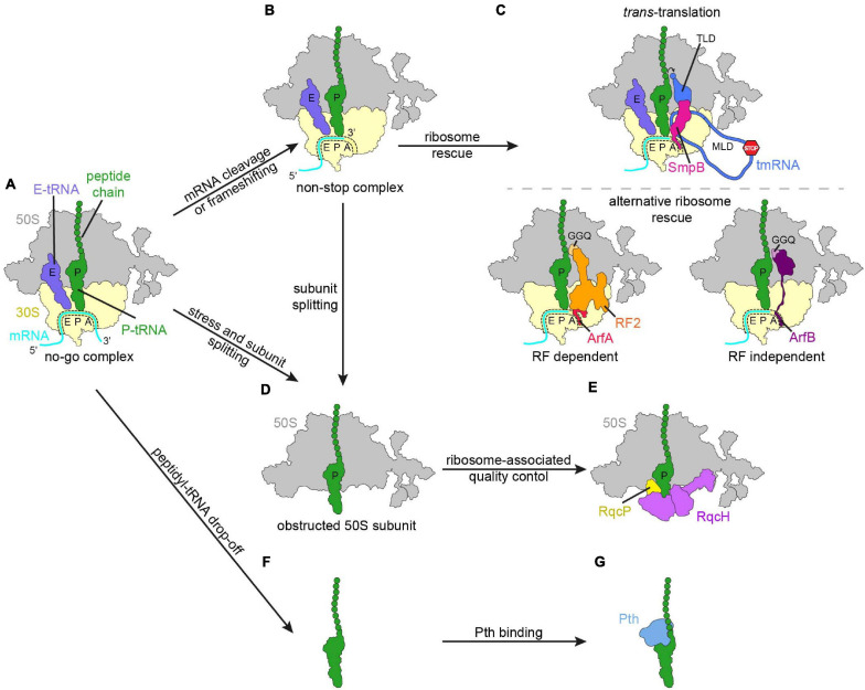
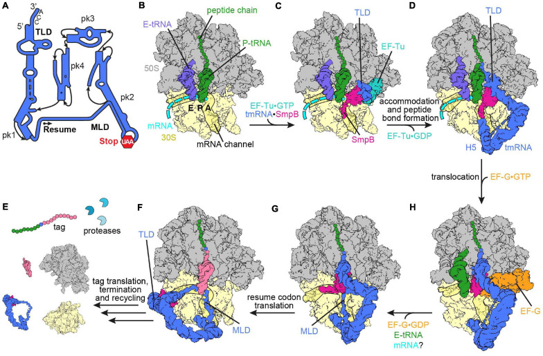
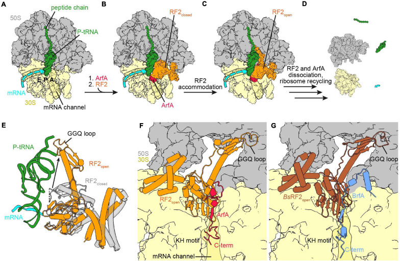
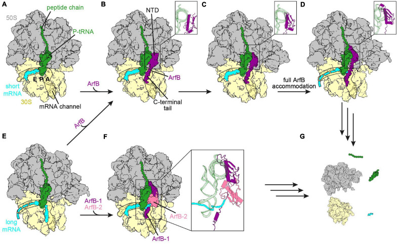
Ribosomes that become stalled on truncated or damaged mRNAs during protein synthesis must be rescued for the cell to survive. Bacteria have evolved a diverse array of rescue pathways to remove the stalled ribosomes from the aberrant mRNA and return them to the free pool of actively translating ribosomes. In addition, some of these pathways target the damaged mRNA and the incomplete nascent polypeptide chain for degradation. This review highlights the recent developments in our mechanistic understanding of bacterial ribosomal rescue systems, including drop-off, trans-translation mediated by transfer-messenger RNA and small protein B, ribosome rescue by the alternative rescue factors ArfA and ArfB, as well as Bacillus ribosome rescue factor A, an additional rescue system found in some Gram-positive bacteria, such as Bacillus subtilis. Finally, we discuss the recent findings of ribosome-associated quality control in particular bacterial lineages mediated by RqcH and RqcP. The importance of rescue pathways for bacterial survival suggests they may represent novel targets for the development of new antimicrobial agents against multi-drug resistant pathogenic bacteria.

Keywords: ArfA; ArfB; RqcH; SmpB; peptidyl-tRNA drop-off; ribosome rescue; ribosome-associated quality control; tmRNA.


Conflict of interest statement

The authors declare that the research was conducted in the absence of any commercial or financial relationships that could be construed as a potential conflict of interest.

Figures

FIGURE 1
FIGURE 1
Schematic overview of regulation mechanisms in response to impeded or aberrant translation. (A) No-go ribosome complex (large subunit, 50S, gray; small subunit, 30S, yellow; mRNA, cyan; P-site tRNA with polypeptide chain, green; E-site tRNA, slate blue). Various responses to the translational pausing generating the no-go complex, like cleavage of the mRNA in the A-site or frameshifting can lead to the ribosome ending up in a non-stop complex (B). (C) The non-stop complex is recognized by ribosome rescue mechanisms. (D,E) In the case that subunit splitting of the no-go complex or the non-stop complex occurred, the obstructed 50S subunit is subjected to bacterial ribosome-associated quality control. (F) Peptidyl-tRNA can drop-off from the ribosome and is recognized by peptidyl-tRNA hydrolase (Pth, blue) (G).
FIGURE 2
FIGURE 2
Ribosome rescue by trans-translation. (A) Schematic representation of tmRNA (pk, pseudoknot; TLD, tRNA-like domain; MLD, mRNA-like domain). The arrow marks the resume codon and the red hexagon indicates the stop codon (UAA) of the MLD. (B) Non-stop complex (large subunit, 50S, gray; small subunit, 30S, yellow) with peptidyl-tRNA (green) in the P-site, E-site tRNA (slate blue) and mRNA (cyan) [PDB ID 4V8Q (Neubauer et al., 2012)]. (C) Delivery of the tmRNA-TLD (blue) in complex with SmpB (violet red) to the non-stop complex by EF-Tu (light sea green) [PDB ID 4V8Q (Neubauer et al., 2012)]. (D) tmRNA⋅SmpB (blue and violet red, respectively) accommodated to the A-site of a non-stop complex [PDB ID 6Q97 (Rae et al., 2019)]. Helix 5 (H5) of tmRNA binds close to the mRNA entry channel. The flexible MLD is indicated by the dashed line. (E) Post-translocation intermediate state of tmRNA⋅SmpB with EF-G (orange) [PDB ID 4V6T (Ramrath et al., 2012)]. tmRNA⋅SmpB and the tRNA (green) are in ap/P and pe/E hybrid states, respectively. (F) Post-translocation complex with tmRNA⋅SmpB in the P-site [PDB ID 6Q98 (Rae et al., 2019)]. The C-terminal tail of SmpB occupies the E-site of the mRNA channel and the resume codon is placed in the A-site. (G) Translation of the resume codon has occurred and the peptide chain was transferred from the TLD to the tRNA (pale violet red) decoding the resume codon [PDB ID 6Q9A (Rae et al., 2019)]. The TLD and SmpB are past the E-site at the outside of the ribosome, while the MLD is fully loaded to the mRNA channel. (H) Translation of the tag peptide, termination at the MLD stop codon and subsequent ribosome recycling have occurred. The tagged peptide is targeted by proteases.
FIGURE 3
FIGURE 3
Ribosome rescue by ArfA and BrfA. (A) Non-stop complex (large subunit, 50S, gray; small subunit, 30S, yellow) with peptide chain and P-site tRNA (both green) and mRNA (cyan) [PDB ID 5MDW (James et al., 2016)]. (B) ArfA (red) binds first to the vacant ribosomal A-site and the mRNA entry channel and recruits RF2 (orange) in the closed conformation to the ribosome [PDB ID 5MDW (James et al., 2016)]. (C) ArfA and RF2 on the non-stop complex after the transition of RF2 to the open conformation [PDB ID 5MGP (Huter et al., 2017c)]. (D) ArfA and RF2 dissociated after release of the peptide chain and ribosome recycling has occurred. (E) Comparison of the RF2 closed [transparent gray, PDB ID 5MDW (James et al., 2016)] and open [orange, PDB ID 5MGP (Huter et al., 2017c)] conformation with P-site tRNA and mRNA for reference. (F,G) View into the A-site with bound ArfA and RF2 [PDB ID 5MGP (Huter et al., 2017c)] (F) or BrfA (blue) and B. subtilis RF2 [brown, PDB ID 6SZS (Shimokawa-Chiba et al., 2019)] (G). Both RF2 are in the open conformation and the C-termini of ArfA and BrfA extend into the mRNA channel, the KH motif and the GGQ-loop are indicated.
FIGURE 4
FIGURE 4
Ribosome rescue by ArfB. (A) Non-stop complex (large subunit, 50S, gray; small subunit, 30S, yellow) with peptidyl-tRNA (green) in the P-site and mRNA extending two nucleotides into the A-site (short mRNA, cyan) [PDB ID 7JSZ (Carbone et al., 2020)]. (B-D) ArfB (purple) bound to the non-stop complex with the NTD in the collapsed (B) [PDB ID 7JSZ (Carbone et al., 2020)], pre-accommodated (C) [PDB ID 7JSW (Carbone et al., 2020)], as well as fully accommodated (D) [PDB ID 6YSS (Chan et al., 2020)] conformation. The conformation of the NTD is enlarged in the inlays. The C-terminal tail extends into the mRNA channel in each conformation. (E) Ribosome with mRNA extending nine nucleotides into the A-site and the mRNA channel (long mRNA, cyan) and peptidyl-tRNA in the P-site [PDB ID 6YSR (Chan et al., 2020)]. (F) ArfB dimer (ArfB, purple and ArfB-2, pale violet red) on the ribosome with the long mRNA sandwiched between the ArfB dimer and the P-site tRNA [PDB ID 7JT1 (Carbone et al., 2020)]. (G) ArfB or the ArfB dimer dissociated and ribosome recycling had occurred.
FIGURE 5
FIGURE 5
Ribosome-associated quality control mediated by RqcH in Bacillus subtilis. (A) Overview of the large subunit (50S, gray) with P-tRNA–nascent chain (green), RqcH (purple), and RqcP (yellow) bound [PDB ID 7AS8 (Crowe-McAuliffe et al., 2021)]. The approximate position of the L1 stalk, which was disordered in this structure, is shown faintly. On the right, a close-up of RqcH, RqcP, and P-tRNA is shown with the domains of RqcH indicated. (B) Proposed mechanism for C-terminal alanine tailing mediated by RqcH and RqcP. For comparison, positions of canonical A-, P-, and E-tRNAs are overlaid in faint gray [PDB ID 6CFJ (Tereshchenkov et al., 2018)]. The large subunit and nascent chain have been omitted for clarity. (i) RqcH (transparent purple) and RqcP (yellow) bound to P-tRNA–nascent chain (green), as in (A). RqcH may also dissociate from this state. (ii) The pre-peptidyl transfer state. The incoming tRNA is positioned similarly to an A-tRNA (salmon) [PDB ID 7AQC (Filbeck et al., 2021)]. (iii) Subsequent to peptidyl transfer and RqcP dissociation, the P-tRNA has moved to the E-site (blue) [State C described in Crowe-McAuliffe et al., 2021). (iv) Similar to (iii), except the E-tRNA has dissociated. Binding of RqcP and movement of RqcH and the L7/L12 stalk completes the cycle [PDB ID 7AS9 (Crowe-McAuliffe et al., 2021)]. (C) The RqcH NFACT-N and HhH domain “decode” tRNAAla(UGC) on the 50S, same as state (B) (ii) [PDB ID 7AQC (Filbeck et al., 2021)]. The tRNA anticodon is splayed in a distorted conformation. (D) Same view as (C), showing conventional interaction of tRNAAla(GGC) with mRNA on a 70S ribosome [PDB ID 6OF6 (Nguyen et al., 2020)] (C,D) were aligned by the tRNA.

References

    1. Abo T., Ueda K., Sunohara T., Ogawa K., Aiba H. (2002). SsrA-mediated protein tagging in the presence of miscoding drugs and its physiological role in Escherichia coli. Genes Cells 7 629–638. 10.1046/j.1365-2443.2002.00549.x - DOI - PubMed
    1. Akabane S., Ueda T., Nierhaus K. H., Takeuchi N. (2014). Ribosome rescue and translation termination at non-standard stop codons by ICT1 in mammalian mitochondria. PLoS Genet. 10:e1004616. 10.1371/journal.pgen.1004616 - DOI - PMC - PubMed
    1. Arenz S., Bock L. V., Graf M., Innis C. A., Beckmann R., Grubmuller H., et al. (2016). A combined cryo-EM and molecular dynamics approach reveals the mechanism of ErmBL-mediated translation arrest. Nat. Commun. 7:12026. 10.1038/ncomms12026 - DOI - PMC - PubMed
    1. Arenz S., Meydan S., Starosta A. L., Berninghausen O., Beckmann R., Vazquez-Laslop N., et al. (2014). Drug sensing by the ribosome induces translational arrest via active site perturbation. Mol. Cell 56 446–452. 10.1016/j.molcel.2014.09.014 - DOI - PMC - PubMed
    1. Aron Z. D., Mehrani A., Hoffer E. D., Connolly K. L., Torhan M. C., Alumasa J. N., et al. (2020). Ribosome rescue inhibitors clear Neisseria gonorrhoeae in vivo using a new mechanism. bioRxiv 10.1101/2020.06.04.132530 - DOI - PMC - PubMed

LinkOut - more resources